 Movie
Movie Controller


+ Open data

- Basic information
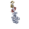
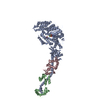
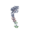
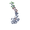
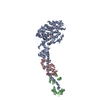
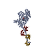
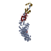
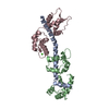
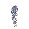
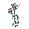
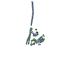
| Entry | Database: PDB / ID: 2w4t | ||||||
|---|---|---|---|---|---|---|---|
| Title | ISOMETRICALLY CONTRACTING INSECT ASYNCHRONOUS FLIGHT MUSCLE | ||||||
 Components Components |
| ||||||
 Keywords Keywords |  CONTRACTILE PROTEIN / NUCLEOTIDE-BINDING / TROPOMYOSIN / LIGHT CHAINS / ACTIN- BINDING / FREEZE SUBSTITUTION / ISOMETRIC CONTRACTION / FREEZING / MICROTOMY / ATP-BINDING / THIN FILAMENT / MOTOR PROTEIN / THICK FILAMENT / MUSCLE PROTEIN / IMAGE PROCESSING / CALMODULIN-BINDING / ACTIN / INSECT / MYOSIN / MUSCLE / TROPONIN / MULTIVARIATE DATA ANALYSIS CONTRACTILE PROTEIN / NUCLEOTIDE-BINDING / TROPOMYOSIN / LIGHT CHAINS / ACTIN- BINDING / FREEZE SUBSTITUTION / ISOMETRIC CONTRACTION / FREEZING / MICROTOMY / ATP-BINDING / THIN FILAMENT / MOTOR PROTEIN / THICK FILAMENT / MUSCLE PROTEIN / IMAGE PROCESSING / CALMODULIN-BINDING / ACTIN / INSECT / MYOSIN / MUSCLE / TROPONIN / MULTIVARIATE DATA ANALYSIS | ||||||
| Function / homology |  Function and homology information Function and homology informationmyosin filament / myosin complex / myofibril / cytoskeletal motor activity / actin filament binding / calmodulin binding / calcium ion binding / ATP bindingSimilarity search - Function | ||||||
| Biological species |  ARGOPECTEN IRRADIANS (bay scallop) ARGOPECTEN IRRADIANS (bay scallop) | ||||||
| Method | ELECTRON MICROSCOPY / helical reconstruction / Resolution: 35 Å | ||||||
 Authors Authors | Wu, S. / Liu, J. / Reedy, M.C. / Tregear, R.T. / Winkler, H. / Franzini-Armstrong, C. / Sasaki, H. / Lucaveche, C. / Goldman, Y.E. / Reedy, M.K. / Taylor, K.A. | ||||||
 Citation Citation | Journal: PLoS One / Year: 2010 Title: Electron tomography of cryofixed, isometrically contracting insect flight muscle reveals novel actin-myosin interactions. Authors: Shenping Wu / Jun Liu / Mary C Reedy / Richard T Tregear / Hanspeter Winkler / Clara Franzini-Armstrong / Hiroyuki Sasaki / Carmen Lucaveche / Yale E Goldman / Michael K Reedy / Kenneth A Taylor /  Abstract: BACKGROUND: Isometric muscle contraction, where force is generated without muscle shortening, is a molecular traffic jam in which the number of actin-attached motors is maximized and all states of ...BACKGROUND: Isometric muscle contraction, where force is generated without muscle shortening, is a molecular traffic jam in which the number of actin-attached motors is maximized and all states of motor action are trapped with consequently high heterogeneity. This heterogeneity is a major limitation to deciphering myosin conformational changes in situ. METHODOLOGY: We used multivariate data analysis to group repeat segments in electron tomograms of isometrically contracting insect flight muscle, mechanically monitored, rapidly frozen, freeze ...METHODOLOGY: We used multivariate data analysis to group repeat segments in electron tomograms of isometrically contracting insect flight muscle, mechanically monitored, rapidly frozen, freeze substituted, and thin sectioned. Improved resolution reveals the helical arrangement of F-actin subunits in the thin filament enabling an atomic model to be built into the thin filament density independent of the myosin. Actin-myosin attachments can now be assigned as weak or strong by their motor domain orientation relative to actin. Myosin attachments were quantified everywhere along the thin filament including troponin. Strong binding myosin attachments are found on only four F-actin subunits, the "target zone", situated exactly midway between successive troponin complexes. They show an axial lever arm range of 77°/12.9 nm. The lever arm azimuthal range of strong binding attachments has a highly skewed, 127° range compared with X-ray crystallographic structures. Two types of weak actin attachments are described. One type, found exclusively in the target zone, appears to represent pre-working-stroke intermediates. The other, which contacts tropomyosin rather than actin, is positioned M-ward of the target zone, i.e. the position toward which thin filaments slide during shortening. CONCLUSION: We present a model for the weak to strong transition in the myosin ATPase cycle that incorporates azimuthal movements of the motor domain on actin. Stress/strain in the S2 domain may ...CONCLUSION: We present a model for the weak to strong transition in the myosin ATPase cycle that incorporates azimuthal movements of the motor domain on actin. Stress/strain in the S2 domain may explain azimuthal lever arm changes in the strong binding attachments. The results support previous conclusions that the weak attachments preceding force generation are very different from strong binding attachments. #1: Journal: J Struct Biol / Year: 2009 Title: Methods for identifying and averaging variable molecular conformations in tomograms of actively contracting insect flight muscle. Authors: Shenping Wu / Jun Liu / Mary C Reedy / Hanspeter Winkler / Michael K Reedy / Kenneth A Taylor / Abstract: During active muscle contraction, tension is generated through many simultaneous, independent interactions between the molecular motor myosin and the actin filaments. The ensemble of myosin motors ...During active muscle contraction, tension is generated through many simultaneous, independent interactions between the molecular motor myosin and the actin filaments. The ensemble of myosin motors displays heterogeneous conformations reflecting different mechanochemical steps of the ATPase pathway. We used electron tomography of actively contracting insect flight muscle fast-frozen, freeze substituted, Araldite embedded, thin-sectioned and stained, to obtain 3D snapshots of the multiplicity of actin-attached myosin structures. We describe procedures for alignment of the repeating lattice of sub-volumes (38.7 nm cross-bridge repeats bounded by troponin) and multivariate data analysis to identify self-similar repeats for computing class averages. Improvements in alignment and classification of repeat sub-volumes reveals (for the first time in active muscle images) the helix of actin subunits in the thin filament and the troponin density with sufficient clarity that a quasiatomic model of the thin filament can be built into the class averages independent of the myosin cross-bridges. We show how quasiatomic model building can identify both strong and weak myosin attachments to actin. We evaluate the accuracy of image classification to enumerate the different types of actin-myosin attachments. | ||||||
| History |
|
- Structure visualization
| Movie |
Movie viewer |
|---|---|
| Structure viewer | Molecule: MolmilJmol/JSmol |
- Downloads & links
-Download
| PDBx/mmCIF format | 2w4t.cif.gz | 7.1 MB | Display | PDBx/mmCIF format |
|---|---|---|---|---|
| PDB format | pdb2w4t.ent.gz | 6.1 MB | Display | PDB format |
| PDBx/mmJSON format | 2w4t.json.gz | Tree view | PDBx/mmJSON format | |
| Others |  Other downloads Other downloads |
-Validation report
| Arichive directory | https://data.pdbj.org/pub/pdb/validation_reports/w4/2w4tftp://data.pdbj.org/pub/pdb/validation_reports/w4/2w4t | HTTPS FTP |
|---|
-Related structure data
| Related structure data |  1561MPC 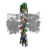 2w49C 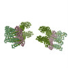 2w4aC 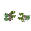 2w4gC 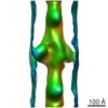 1584F M: map data used to model this data P: unfit; worng pairing?*YM C: citing same article ( F: fitted*YM |
|---|---|
| Similar structure data |

-Links
 PDBj
PDBj


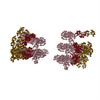
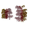
- Assembly
| Deposited unit | 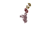
|
|---|---|
| 1 | 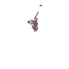
|
| 2 | 
|
| 3 | 
|
| Number of models | 43 |
-Components
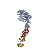
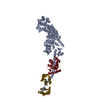
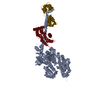
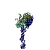
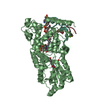
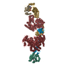
| #1: Protein | Myosin / MYOSIN S1 Mass: 94843.883 Da / Num. of mol.: 1 / Fragment: RESIDUES 5-835 / Source method: isolated from a natural source / Details: MYOSIN S1 HEAVY CHAIN / Source: (natural) ARGOPECTEN IRRADIANS (bay scallop) / References: UniProt: P24733 |
|---|---|
| #2: Protein | Mass: 15575.700 Da / Num. of mol.: 1 / Fragment: RESIDUES 16-151 / Source method: isolated from a natural source Details: MYOSIN REGULATORY LIGHT CHAIN, RLC, STRIATED ADDUCTOR MUSCLE Source: (natural) ARGOPECTEN IRRADIANS (bay scallop) / References: UniProt: P13543 |
| #3: Protein | Mass: 17051.912 Da / Num. of mol.: 1 / Fragment: RESIDUES 5-155 / Source method: isolated from a natural source Details: MYOSIN ESSENTIAL LIGHT CHAIN, ELC, STRIATED ADDUCTOR MUSCLE Source: (natural) ARGOPECTEN IRRADIANS (bay scallop) / References: UniProt: P07291 |
-Experimental details
-Experiment
| Experiment | Method: ELECTRON MICROSCOPY |
|---|---|
| EM experiment | Aggregation state: FILAMENT / 3D reconstruction method: helical reconstruction |
- Sample preparation
| Component | Name: ISOMETRICALLY CONTRACTING ASYNCHRONOUS INSECT FLIGHT MUSCLE FROM THE LARGE WATERBUG LETHOCERUS INDICUS Type: COMPLEX Details: TOMOGRAPHIC TILT SERIES COLLECTED AROUND TWO MUTUALLY ORTHOGONAL TILT AXES USING THE SAXTON SCHEME FOR DETERMINING THE TILT ANGLES. THE TWO TILT SERIES WERE MERGED USING MARKER FREE ...Details: TOMOGRAPHIC TILT SERIES COLLECTED AROUND TWO MUTUALLY ORTHOGONAL TILT AXES USING THE SAXTON SCHEME FOR DETERMINING THE TILT ANGLES. THE TWO TILT SERIES WERE MERGED USING MARKER FREE ALIGNMENT AND THE TWO INDEPENDENT TILT SERIES COMBINED USING IMOD. |
|---|---|
| Buffer solution | Name: 20 MM MOPS BUFFER, 5 MM NAN3, AND MGCL2, ATP, CACL2, AND EGTA IN VARYING MILLIMOLAR MILLIMOLAR CONCENTRATIONS Details: 20 MM MOPS BUFFER, 5 MM NAN3, AND MGCL2, ATP, CACL2, AND EGTA IN VARYING MILLIMOLAR MILLIMOLAR CONCENTRATIONS |
| Specimen | Embedding applied: YES / Shadowing applied: NO / Staining applied: NO / Vitrification applied: NO |
| Specimen support | Details: CARBON |
| Vitrification | Instrument: HOMEMADE PLUNGER / Cryogen name: HELIUM |
- Electron microscopy imaging
| Microscopy | Model: FEI/PHILIPS CM300FEG/T / Details: NONE |
|---|---|
| Electron gun | Electron source: FIELD EMISSION GUN / Accelerating voltage: 300 kV / Illumination mode: FLOOD BEAM |
| Electron lens | Mode: BRIGHT FIELDBright-field microscopy / Cs: 2 mm |
| Specimen holder | Tilt angle max: 72 ° / Tilt angle min: -72 ° |
| Image recording | Film or detector model: TVIPS TEMCAM-F224 (2k x 2k) |
| Radiation wavelength | Relative weight: 1 |
- Processing
| 3D reconstruction | Resolution method: FSC 0.5 CUT-OFF Details: NOTE THAT OUR LOWEST RESOLUTION DATA IS AT INVERSE 1 MICRON. NUMBER OF FOURIER COEFFICIENTS IS ALMOST A HALF MILLION. THESE COORDINATES WERE FITTED TO AVERAGED SUBVOLUMES OBTAINED FROM A ...Details: NOTE THAT OUR LOWEST RESOLUTION DATA IS AT INVERSE 1 MICRON. NUMBER OF FOURIER COEFFICIENTS IS ALMOST A HALF MILLION. THESE COORDINATES WERE FITTED TO AVERAGED SUBVOLUMES OBTAINED FROM A DUAL AXIS TOMOGRAM. THE FITTING WAS DONE MANUALLY USING THE CRYSTALLOGRAPHIC MODEL FITTING PROGRAM O. THERE ARE 43 MODELS. Symmetry type: HELICAL | ||||||||||||
|---|---|---|---|---|---|---|---|---|---|---|---|---|---|
| Refinement | Highest resolution: 35 Å | ||||||||||||
| Refinement step | Cycle: LAST / Highest resolution: 35 Å
|
