 Movie
Movie Controller


+ Open data

- Basic information
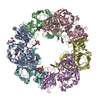
| Entry | Database: PDB / ID: 2rec | ||||||
|---|---|---|---|---|---|---|---|
| Title | RECA HEXAMER MODEL, ELECTRON MICROSCOPY | ||||||
 Components Components | RECA | ||||||
 Keywords Keywords | HELICASE / DNA DAMAGE / DNA RECOMBINATION / SOS RESPONSE / ATP-BINDING / DNA-BINDING | ||||||
| Function / homology |  Function and homology informationDNA polymerase V complex / homologous recombination / recombinational repair / SOS response / ATP-dependent DNA damage sensor activity / response to ionizing radiation / translesion synthesis / ATP-dependent activity, acting on DNA / cell motility / single-stranded DNA binding ...DNA polymerase V complex / homologous recombination / recombinational repair / SOS response / ATP-dependent DNA damage sensor activity / response to ionizing radiation / translesion synthesis / ATP-dependent activity, acting on DNA / cell motility / single-stranded DNA binding / DNA-binding transcription factor binding / DNA recombination / damaged DNA binding / DNA damage response / ATP hydrolysis activity / ATP binding / cytoplasm Function and homology informationDNA polymerase V complex / homologous recombination / recombinational repair / SOS response / ATP-dependent DNA damage sensor activity / response to ionizing radiation / translesion synthesis / ATP-dependent activity, acting on DNA / cell motility / single-stranded DNA binding ...DNA polymerase V complex / homologous recombination / recombinational repair / SOS response / ATP-dependent DNA damage sensor activity / response to ionizing radiation / translesion synthesis / ATP-dependent activity, acting on DNA / cell motility / single-stranded DNA binding / DNA-binding transcription factor binding / DNA recombination / damaged DNA binding / DNA damage response / ATP hydrolysis activity / ATP binding / cytoplasmSimilarity search - Function | ||||||
| Biological species |  Escherichia coli (E. coli) Escherichia coli (E. coli) | ||||||
| Method | ELECTRON MICROSCOPY / helical reconstruction / negative staining | ||||||
 Authors Authors | Yu, X. / Egelman, E.H. | ||||||
 Citation Citation | Journal: Nat Struct Biol / Year: 1997 Title: The RecA hexamer is a structural homologue of ring helicases. Abstract: The RecA protein forms a hexameric ring that is similar to the core of the F1-ATPase. Several lines of evidence suggest that this hexamer may be a structural homologue of ring helicases. | ||||||
| History |
|
- Structure visualization
| Movie |
Movie viewer |
|---|---|
| Structure viewer | Molecule: MolmilJmol/JSmol |
- Downloads & links
-Download
| PDBx/mmCIF format | 2rec.cif.gz | 55.6 KB | Display | PDBx/mmCIF format |
|---|---|---|---|---|
| PDB format | pdb2rec.ent.gz | 34.9 KB | Display | PDB format |
| PDBx/mmJSON format | 2rec.json.gz | Tree view | PDBx/mmJSON format | |
| Others |  Other downloads Other downloads |
-Validation report
| Arichive directory | https://data.pdbj.org/pub/pdb/validation_reports/re/2recftp://data.pdbj.org/pub/pdb/validation_reports/re/2rec | HTTPS FTP |
|---|
-Related structure data
| Similar structure data |
|---|


-Links
 PDBj
PDBj
- Assembly
| Deposited unit | 
|
|---|---|
| 1 |
|
-Components
| #1: Protein | / Coordinate model: Cα atoms only Mass: 38016.277 Da / Num. of mol.: 6 / Source method: isolated from a natural source / Source: (natural) Escherichia coli (E. coli) / References: UniProt: P0A7G6 |
|---|
-Experimental details
-Experiment
| Experiment | Method: ELECTRON MICROSCOPY |
|---|---|
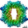
| EM experiment | Aggregation state: FILAMENT / 3D reconstruction method: helical reconstruction |
- Sample preparation
| Component | Name: RecA filament / Type: COMPLEX |
|---|---|
| Specimen | Embedding applied: NO / Shadowing applied: NO / Staining applied: YES / Vitrification applied: NO |
| EM staining | Type: NEGATIVE / Material: uranyl acetate |
| Crystal grow | *PLUS Method: other |
- Electron microscopy imaging
| Microscopy | Model: JEOL 1200EXII |
|---|---|
| Electron gun | Accelerating voltage: 80 kV / Illumination mode: FLOOD BEAM |
| Electron lens | Mode: BRIGHT FIELDBright-field microscopy |
| Detector | Date: Sep 1, 1996 |
- Processing
| Software | Name: XTALVIEW / Classification: refinement | ||||||||||||
|---|---|---|---|---|---|---|---|---|---|---|---|---|---|
| Refinement step | Cycle: LAST
|
