Movie
Movie Controller

[English] 日本語
 Yorodumi
- PDB-2r6p: Fit of E protein and Fab 1A1D-2 into 24 angstrom resolution cryoE... -
+ Open data

- Basic information
| Entry | Database: PDB / ID: 2r6p | ||||||
|---|---|---|---|---|---|---|---|
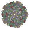
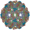
| Title | Fit of E protein and Fab 1A1D-2 into 24 angstrom resolution cryoEM map of Fab complexed with dengue 2 virus. | ||||||
 Components Components |
| ||||||
 Keywords Keywords | Virus/Immune System /  Fab / dengue / virus / neutralization / Virus-Immune System COMPLEX / icosahedral virus Fab / dengue / virus / neutralization / Virus-Immune System COMPLEX / icosahedral virus | ||||||
| Function / homology |  Function and homology information Function and homology informationclathrin-dependent endocytosis of virus by host cell / viral nucleocapsid / host cell endoplasmic reticulum membrane / protein dimerization activity / fusion of virus membrane with host endosome membrane / viral envelope / virion attachment to host cell / virion membrane / extracellular region / membraneSimilarity search - Function | ||||||
| Biological species |  Dengue virus 2 Puerto Rico/PR159-S1/1969 Dengue virus 2 Puerto Rico/PR159-S1/1969 Mus musculus (house mouse) Mus musculus (house mouse) | ||||||
| Method | ELECTRON MICROSCOPY / single particle reconstruction / cryo EM / Resolution: 24 Å | ||||||
 Authors Authors | Lok, S.M. / Kostyuchenko, V.K. / Holdaway, H.A. / Chipman, P.R. / Kuhn, R.J. / Rossmann, M.G. | ||||||
 Citation Citation | Journal: Nat Struct Mol Biol / Year: 2008 Title: Binding of a neutralizing antibody to dengue virus alters the arrangement of surface glycoproteins. Authors: Shee-Mei Lok / Victor Kostyuchenko / Grant E Nybakken / Heather A Holdaway / Anthony J Battisti / Soila Sukupolvi-Petty / Dagmar Sedlak / Daved H Fremont / Paul R Chipman / John T Roehrig / ...Authors: Shee-Mei Lok / Victor Kostyuchenko / Grant E Nybakken / Heather A Holdaway / Anthony J Battisti / Soila Sukupolvi-Petty / Dagmar Sedlak / Daved H Fremont / Paul R Chipman / John T Roehrig / Michael S Diamond / Richard J Kuhn / Michael G Rossmann /  Abstract: The monoclonal antibody 1A1D-2 has been shown to strongly neutralize dengue virus serotypes 1, 2 and 3, primarily by inhibiting attachment to host cells. A crystal structure of its antigen binding ...The monoclonal antibody 1A1D-2 has been shown to strongly neutralize dengue virus serotypes 1, 2 and 3, primarily by inhibiting attachment to host cells. A crystal structure of its antigen binding fragment (Fab) complexed with domain III of the viral envelope glycoprotein, E, showed that the epitope would be partially occluded in the known structure of the mature dengue virus. Nevertheless, antibody could bind to the virus at 37 degrees C, suggesting that the virus is in dynamic motion making hidden epitopes briefly available. A cryo-electron microscope image reconstruction of the virus:Fab complex showed large changes in the organization of the E protein that exposed the epitopes on two of the three E molecules in each of the 60 icosahedral asymmetric units of the virus. The changes in the structure of the viral surface are presumably responsible for inhibiting attachment to cells. | ||||||
| History |
|
- Structure visualization
| Movie |
Movie viewer |
|---|---|
| Structure viewer | Molecule: MolmilJmol/JSmol |
- Downloads & links
-Download
| PDBx/mmCIF format | 2r6p.cif.gz | 76.5 KB | Display | PDBx/mmCIF format |
|---|---|---|---|---|
| PDB format | pdb2r6p.ent.gz | 47 KB | Display | PDB format |
| PDBx/mmJSON format | 2r6p.json.gz | Tree view | PDBx/mmJSON format | |
| Others |  Other downloads Other downloads |
-Validation report
| Arichive directory | https://data.pdbj.org/pub/pdb/validation_reports/r6/2r6pftp://data.pdbj.org/pub/pdb/validation_reports/r6/2r6p | HTTPS FTP |
|---|
-Related structure data
| Related structure data | 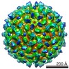 1418MC 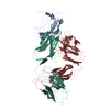 2r29C 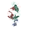 2r69C M: map data used to model this data C: citing same article ( |
|---|---|
| Similar structure data |


-Links
 PDBj
PDBj


- Assembly
| Deposited unit | 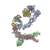
|
|---|---|
| 1 | x 60
|
| 2 |
|
| 3 | x 5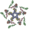
|
| 4 | x 6
|
| 5 | 
|
| Symmetry | Point symmetry: (Schoenflies symbol: I (icosahedral)) |
-Components
| #1: Protein | Mass: 43403.984 Da / Num. of mol.: 3 / Fragment: E protein Source method: isolated from a genetically manipulated source Source: (gene. exp.) Dengue virus 2 Puerto Rico/PR159-S1/1969Genus: Flavivirus / Species: Dengue virus / Strain: PR-159-S1 / Gene: E protein / Plasmid: pET21 / Species (production host): Escherichia coli / Production host:  Escherichia coli BL21(DE3) (bacteria) / Strain (production host): BL21(DE3) / References: UniProt: P18356, UniProt: Q66394*PLUS Escherichia coli BL21(DE3) (bacteria) / Strain (production host): BL21(DE3) / References: UniProt: P18356, UniProt: Q66394*PLUS#2: Antibody | Mass: 23068.715 Da / Num. of mol.: 2 Source method: isolated from a genetically manipulated source Source: (gene. exp.) Mus musculus (house mouse) / Strain: Balbc / Gene: immunoglobulin / Cell line (production host): hybridoma / References: UniProt: Q6PJB2*PLUS#3: Antibody | Mass: 22808.279 Da / Num. of mol.: 2 Source method: isolated from a genetically manipulated source Source: (gene. exp.) Mus musculus (house mouse) / Strain: balbc / Gene: immunoglobulin / Cell line (production host): hybridoma |
|---|
-Experimental details
-Experiment
| Experiment | Method: ELECTRON MICROSCOPY |
|---|---|
| EM experiment | Aggregation state: PARTICLE / 3D reconstruction method: single particle reconstruction |
- Sample preparation
| Component | Name: Fab Fragment of MAb 1A1D-2 complexed with Dengue 2 virus Type: VIRUS / Details: 12 mM Tris-HCl, 120 mM NaCl, 1 mM EDTA |
|---|---|
| Details of virus | Host category: MOSQUITO / Type: VIRION |
| Natural host | Organism: Aedes albopictus / Strain: C6/36 cells |
| Buffer solution | pH: 7.6 |
| Specimen | Conc.: 0.6 mg/ml / Embedding applied: NO / Shadowing applied: NO / Staining applied: NO / Vitrification applied: YES |
| Specimen support | Details: 400 mesh copper grid |
| Vitrification | Instrument: HOMEMADE PLUNGER / Cryogen name: ETHANE Details: SAMPLES WERE PREPARED AS THIN LAYERS OF VITREOUS ICE AND MAINTAINED AT LIQUID NITROGEN TEMPERATURE IN THE ELECTRON MICROSCOPE |
- Electron microscopy imaging
| Microscopy | Model: FEI/PHILIPS CM200T / Date: Mar 21, 2007 / Details: low dose |
|---|---|
| Electron gun | Electron source: FIELD EMISSION GUN / Accelerating voltage: 200 kV / Illumination mode: FLOOD BEAM |
| Electron lens | Mode: BRIGHT FIELDBright-field microscopy / Nominal magnification: 50000 X / Calibrated magnification: 51040 X / Nominal defocus max: 3373 nm / Nominal defocus min: 2276 nm / Cs: 2 mm |
| Specimen holder | Temperature: 87 K / Tilt angle max: 0 ° / Tilt angle min: 0 ° |
| Image recording | Electron dose: 2.39 e/Å2 / Film or detector model: KODAK SO-163 FILM |
| Radiation | Protocol: SINGLE WAVELENGTH / Monochromatic (M) / Laue (L): M / Scattering type: x-ray |
| Radiation wavelength | Relative weight: 1 |
- Processing
| EM software |
| ||||||||||||
|---|---|---|---|---|---|---|---|---|---|---|---|---|---|
| CTF correction | Details: phase flip | ||||||||||||
| Symmetry | Point symmetry: I (icosahedral) | ||||||||||||
| 3D reconstruction | Method: projection matching / Resolution: 24 Å / Num. of particles: 2885 / Actual pixel size: 2.74 Å / Details: THE COORDINATES IN THIS ENTRY CONTAIN CA ONLY / Symmetry type: POINT | ||||||||||||
| Atomic model building | Protocol: RIGID BODY FIT / Space: REAL / Target criteria: Using EMFIT Details: METHOD--place coordinates manually and then optimise position using program REFINEMENT PROTOCOL--rigid body | ||||||||||||
| Atomic model building | PDB-ID: 1THD Accession code: 1THD / Source name: PDB / Type: experimental model | ||||||||||||
| Refinement step | Cycle: LAST
|