 Movie
Movie Controller

[English] 日本語
 Yorodumi
- PDB-2h50: Multiple distinct assemblies reveal conformational flexibility in... -
+ Open data

- Basic information
| Entry | Database: PDB / ID: 2h50 | ||||||
|---|---|---|---|---|---|---|---|
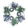
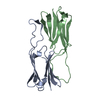
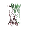
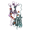
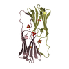
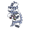
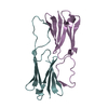
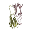
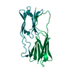
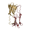
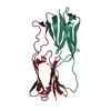
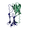
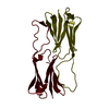
| Title | Multiple distinct assemblies reveal conformational flexibility in the small heat shock protein Hsp26 | ||||||
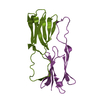
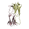
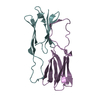
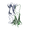
 Components Components | small heat shock protein Hsp26 | ||||||
 Keywords Keywords | CHAPERONE / alpha-crystallin / chaperones / heat shock proteins / single particle reconstruction | ||||||
| Function / homology |  Function and homology information Function and homology informationresponse to salt stress / response to hydrogen peroxide / protein homooligomerization / unfolded protein binding / protein folding / protein complex oligomerization / response to heat / cytoplasm Similarity search - Function | ||||||
| Biological species |  | ||||||
| Method | ELECTRON MICROSCOPY / single particle reconstruction / cryo EM / Resolution: 10.8 Å | ||||||
 Authors Authors | White, H.E. / Orlova, E.V. / Chen, S. / Wang, L. / Ignatiou, A. / Gowen, B. / Stromer, T. / Franzmann, T.M. / Haslbeck, M. / Buchner, J. / Saibil, H.R. | ||||||
 Citation Citation | Journal: Structure / Year: 2006 Title: Multiple distinct assemblies reveal conformational flexibility in the small heat shock protein Hsp26. Authors: Helen E White / Elena V Orlova / Shaoxia Chen / Luchun Wang / Athanasios Ignatiou / Brent Gowen / Thusnelda Stromer / Titus M Franzmann / Martin Haslbeck / Johannes Buchner / Helen R Saibil /  Abstract: Small heat shock proteins are a superfamily of molecular chaperones that suppress protein aggregation and provide protection from cell stress. A key issue for understanding their action is to define ...Small heat shock proteins are a superfamily of molecular chaperones that suppress protein aggregation and provide protection from cell stress. A key issue for understanding their action is to define the interactions of subunit domains in these oligomeric assemblies. Cryo-electron microscopy of yeast Hsp26 reveals two distinct forms, each comprising 24 subunits arranged in a porous shell with tetrahedral symmetry. The subunits form elongated, asymmetric dimers that assemble via trimeric contacts. Modifications of both termini cause rearrangements that yield a further four assemblies. Each subunit contains an N-terminal region, a globular middle domain, the alpha-crystallin domain, and a C-terminal tail. Twelve of the C termini form 3-fold assembly contacts which are inserted into the interior of the shell, while the other 12 C termini form contacts on the surface. Hinge points between the domains allow a variety of assembly contacts, providing the flexibility required for formation of supercomplexes with non-native proteins. | ||||||
| History |
| ||||||
| Remark 999 | SEQUENCE THE SEQUENCE LISTED HERE IS THAT OF HEAT SHOCK PROTEIN 16.9B CORRESPONDING TO THE PDB ...SEQUENCE THE SEQUENCE LISTED HERE IS THAT OF HEAT SHOCK PROTEIN 16.9B CORRESPONDING TO THE PDB ENTRY 1GME. THE ACTUAL SEQUENCE IN THE SAMPLE IS THAT OF UNIPROT ENTRY P15992. |
- Structure visualization
| Movie |
Movie viewer |
|---|---|
| Structure viewer | Molecule: MolmilJmol/JSmol |
- Downloads & links
-Download
| PDBx/mmCIF format | 2h50.cif.gz | 427.9 KB | Display | PDBx/mmCIF format |
|---|---|---|---|---|
| PDB format | pdb2h50.ent.gz | 359.8 KB | Display | PDB format |
| PDBx/mmJSON format | 2h50.json.gz | Tree view | PDBx/mmJSON format | |
| Others |  Other downloads Other downloads |
-Validation report
| Summary document | 2h50_validation.pdf.gz | 882.5 KB | Display | wwPDB validaton report |
|---|---|---|---|---|
| Full document | 2h50_full_validation.pdf.gz | 937.7 KB | Display | |
| Data in XML | 2h50_validation.xml.gz | 62 KB | Display | |
| Data in CIF | 2h50_validation.cif.gz | 74.8 KB | Display | |
| Arichive directory | https://data.pdbj.org/pub/pdb/validation_reports/h5/2h50ftp://data.pdbj.org/pub/pdb/validation_reports/h5/2h50 | HTTPS FTP |
-Related structure data
| Related structure data | 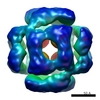 1221MC 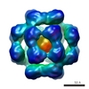 1226C 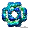 1227C 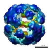 1228C 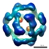 1229C 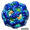 1230C  2h53C C: citing same article ( M: map data used to model this data |
|---|---|
| Similar structure data |


-Links
 PDBj
PDBj


- Assembly


-Components
| #1: Protein | Mass: 10680.172 Da / Num. of mol.: 24 / Source method: isolated from a natural source / Source: (natural) Sequence details | EXPERIMENT | |
|---|
-Experimental details
-Experiment
| Experiment | Method: ELECTRON MICROSCOPY |
|---|---|
| EM experiment | Aggregation state: PARTICLE / 3D reconstruction method: single particle reconstruction |
- Sample preparation
| Component | Name: small heat shock protein Hsp26 complex / Type: COMPLEX / Details: 24-mer |
|---|---|
| Buffer solution | Name: 40 mM Hepes, 50 mM NaCl, 2 mM EDTA, 1 mM DTT / pH: 7.4 / Details: 40 mM Hepes, 50 mM NaCl, 2 mM EDTA, 1 mM DTT |
| Specimen | Conc.: 1 mg/ml / Embedding applied: NO / Shadowing applied: NO / Staining applied: NO / Vitrification applied: YES |
| Vitrification | Cryogen name: ETHANE |
- Electron microscopy imaging
| Experimental equipment |  Model: Tecnai F20 / Image courtesy: FEI Company |
|---|---|
| Microscopy | Model: FEI TECNAI F20 |
| Electron gun | Electron source:  FIELD EMISSION GUN / Accelerating voltage: 200 kV / Illumination mode: FLOOD BEAM FIELD EMISSION GUN / Accelerating voltage: 200 kV / Illumination mode: FLOOD BEAM |
| Electron lens | Mode: BRIGHT FIELD / Nominal magnification: 38000 X / Nominal defocus max: 3400 nm / Nominal defocus min: 1700 nm |
| Image recording | Film or detector model: KODAK SO-163 FILM |
| Radiation | Protocol: SINGLE WAVELENGTH / Monochromatic (M) / Laue (L): M / Scattering type: x-ray |
| Radiation wavelength | Relative weight: 1 |
- Processing
| EM software |
| ||||||||||||
|---|---|---|---|---|---|---|---|---|---|---|---|---|---|
| CTF correction | Details: phase flipping | ||||||||||||
| Symmetry | Point symmetry: T (tetrahedral) | ||||||||||||
| 3D reconstruction | Method: ANGULAR RECONSTITUTION / Resolution: 10.8 Å / Resolution method: FSC 0.5 CUT-OFF / Num. of particles: 10000 / Symmetry type: POINT | ||||||||||||
| Atomic model building | Protocol: RIGID BODY FIT / Space: RECIPROCAL / Target criteria: best visual fit using the program O / Details: REFINEMENT PROTOCOL--rigid body | ||||||||||||
| Atomic model building | PDB-ID: 1GME Accession code: 1GME / Source name: PDB / Type: experimental model | ||||||||||||
| Refinement step | Cycle: LAST
|
