 Movie
Movie Controller

[English] 日本語
 Yorodumi
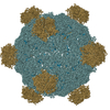
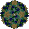
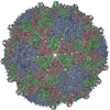
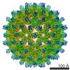
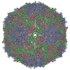
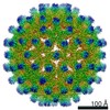
- PDB-1m0f: Structural Studies of Bacteriophage alpha3 Assembly, Cryo-electro... -
+ Open data

- Basic information
| Entry | Database: PDB / ID: 1m0f | ||||||
|---|---|---|---|---|---|---|---|
| Title | Structural Studies of Bacteriophage alpha3 Assembly, Cryo-electron microscopy | ||||||
 Components Components |
| ||||||
 Keywords Keywords |  VIRUS / Bacteriophage / Cryo Electron Microscopy / Procapsid / morphogenesis / microviridae / assembly / Icosahedral virus VIRUS / Bacteriophage / Cryo Electron Microscopy / Procapsid / morphogenesis / microviridae / assembly / Icosahedral virus | ||||||
| Function / homology |  Function and homology information Function and homology informationmodulation by virus of host process / viral procapsid maturation / T=1 icosahedral viral capsid / viral capsid / host cell cytoplasm / symbiont entry into host cell / virion attachment to host cell / structural molecule activitySimilarity search - Function | ||||||
| Biological species |  Enterobacteria phage alpha3 (virus) Enterobacteria phage alpha3 (virus) | ||||||
| Method | ELECTRON MICROSCOPY / single particle reconstruction / cryo EM / Resolution: 16 Å | ||||||
 Authors Authors | Bernal, R.A. / Hafenstein, S. / Olson, N.H. / Bowman, V.D. / Chipman, P.R. / Baker, T.S. / Fane, B.A. / Rossmann, M.G. | ||||||
 Citation Citation | Journal: J Mol Biol / Year: 2003 Title: Structural studies of bacteriophage alpha3 assembly. Authors: Ricardo A Bernal / Susan Hafenstein / Norman H Olson / Valorie D Bowman / Paul R Chipman / Timothy S Baker / Bentley A Fane / Michael G Rossmann /  Abstract: Bacteriophage alpha3 is a member of the Microviridae, a family of small, single-stranded, icosahedral phages that include phiX174. These viruses have an ssDNA genome associated with approximately 12 ...Bacteriophage alpha3 is a member of the Microviridae, a family of small, single-stranded, icosahedral phages that include phiX174. These viruses have an ssDNA genome associated with approximately 12 copies of an H pilot protein and 60 copies of a small J DNA-binding protein. The surrounding capsid consists of 60 F coat proteins decorated with 12 pentameric spikes of G protein. Assembly proceeds via a 108S empty procapsid that requires the external D and internal B scaffolding proteins for its formation. The alpha3 "open" procapsid structural intermediate was determined to 15A resolution by cryo-electron microscopy (cryo-EM). Unlike the phiX174 "closed" procapsid and the infectious virion, the alpha3 open procapsid has 30A wide pores at the 3-fold vertices and 20A wide gaps between F pentamers as a result of the disordering of two helices in the F capsid protein. The large pores are probably used for DNA entry and internal scaffolding protein exit during DNA packaging. Portions of the B scaffolding protein are located at the 5-fold axes under the spike and in the hydrophobic pocket on the inner surface of the capsid. Protein B appears to have autoproteolytic activity that cleaves at an Arg-Phe motif and probably facilitates the removal of the protein through the 30A wide pores. The structure of the alpha3 mature virion was solved to 3.5A resolution by X-ray crystallography and was used to interpret the open procapsid cryo-EM structure. The main differences between the alpha3 and phiX174 virion structures are in the spike and the DNA-binding proteins. The alpha3 pentameric spikes have a rotation of 3.5 degrees compared to those of phiX174. The alpha3 DNA-binding protein, which is shorter by 13 amino acid residues at its amino end when compared to the phiX174 J protein, retains its carboxy-terminal-binding site on the internal surface of the capsid protein. The icosahedrally ordered structural component of the ssDNA appears to be substantially increased in alpha3 compared to phiX174, allowing the building of about 10% of the ribose-phosphate backbone. | ||||||
| History |
| ||||||
| Remark 999 | SEQUENCE Residue 160 of the F protein is an ARG according to the reported sequence but no density ...SEQUENCE Residue 160 of the F protein is an ARG according to the reported sequence but no density is seen for the side chain in the crystal structure for this residue. After a structural sequence alignment with homologous bacteriophages phix174 and G4, residue 160 was found to be a glycine in the other phages. Consequently, the authors state residue 160 should be a Glycine. CHAINS 1,2,3,4 ARE SCAFFOLDING PROTEIN D AND CHAIN B IS SCAFFOLDING PROTEIN B, ALL FROM BACTERIOPHAGE ALPHA3. THE PROTEIN SEQUENCES WERE TAKEN FROM 1CD3 PHIX174 COORDINATES AND FITTED INTO THE CRYO-EM STRUCTURE. THE SEQUENCE DATABASE REFERENCE FOR THE PHIX174 SCAFFOLDING PROTEINS D AND B ARE P03637 AND P07929 RESPECTIVELY. |
- Structure visualization
| Movie |
Movie viewer |
|---|---|
| Structure viewer | Molecule: MolmilJmol/JSmol |
- Downloads & links
-Download
| PDBx/mmCIF format | 1m0f.cif.gz | 51.3 KB | Display | PDBx/mmCIF format |
|---|---|---|---|---|
| PDB format | pdb1m0f.ent.gz | 28 KB | Display | PDB format |
| PDBx/mmJSON format | 1m0f.json.gz | Tree view | PDBx/mmJSON format | |
| Others |  Other downloads Other downloads |
-Validation report
| Arichive directory | https://data.pdbj.org/pub/pdb/validation_reports/m0/1m0fftp://data.pdbj.org/pub/pdb/validation_reports/m0/1m0f | HTTPS FTP |
|---|
-Related structure data


-Links
 PDBj
PDBj


- Assembly
| Deposited unit | 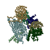
|
|---|---|
| 1 | x 60
|
| 2 |
|
| 3 | x 5
|
| 4 | x 6
|
| 5 | 
|
| Symmetry | Point symmetry: (Hermann–Mauguin notation: 532 / Schoenflies symbol: I (icosahedral)) |
-Components
| #1: Protein | / GPD / Coordinate model: Cα atoms only Mass: 16953.316 Da / Num. of mol.: 4 Source method: isolated from a genetically manipulated source Source: (gene. exp.) Enterobacteria phage alpha3 (virus) / Genus: Microvirus / Production host:  Escherichia coli (E. coli) / References: UniProt: P69486*PLUS Escherichia coli (E. coli) / References: UniProt: P69486*PLUS#2: Protein | | / F protein / GPF / Coordinate model: Cα atoms onlyMass: 49294.277 Da / Num. of mol.: 1 Source method: isolated from a genetically manipulated source Source: (gene. exp.) Enterobacteria phage alpha3 (virus) / Genus: Microvirus / Production host: Escherichia coli (E. coli) / References: UniProt: P08767#3: Protein | | Mass: 19598.990 Da / Num. of mol.: 1 Source method: isolated from a genetically manipulated source Source: (gene. exp.) Enterobacteria phage alpha3 (virus) / Genus: Microvirus / Production host: Escherichia coli (E. coli) / References: UniProt: P31281#4: Protein | | / GPB / Coordinate model: Cα atoms onlyMass: 8150.042 Da / Num. of mol.: 1 Source method: isolated from a genetically manipulated source Source: (gene. exp.) Enterobacteria phage alpha3 (virus) / Genus: Microvirus / Production host: Escherichia coli (E. coli) |
|---|
-Experimental details
-Experiment
| Experiment | Method: ELECTRON MICROSCOPY |
|---|---|
| EM experiment | Aggregation state: PARTICLE / 3D reconstruction method: single particle reconstruction |
- Sample preparation
| Component | Name: Procapsid of the bacteriophage alpha3 / Type: VIRUS |
|---|---|
| Details of virus | Details: Assembly intermediate called the Procapsid / Host category: BACTERIA(EUBACTERIA) / Isolate: SPECIES |
| Natural host | Organism: Escherichia coli / Strain: slyD |
| Buffer solution | Name: 10 mM Tris and 1 mM EDTA / pH: 7.5 / Details: 10 mM Tris and 1 mM EDTA |
| Specimen | Conc.: 8 mg/ml / Embedding applied: NO / Shadowing applied: NO / Staining applied: NO / Vitrification applied: YES |
| Crystal grow | *PLUS Method: other / Details: cryo-electron microscopy |
- Electron microscopy imaging
| Microscopy | Model: FEI/PHILIPS CM200FEG/ST / Date: Oct 16, 1998 |
|---|---|
| Electron gun | Electron source: FIELD EMISSION GUN / Accelerating voltage: 200 kV / Illumination mode: FLOOD BEAM |
| Electron lens | Mode: BRIGHT FIELDBright-field microscopy / Nominal magnification: 38000 X / Calibrated magnification: 40000 X / Nominal defocus max: 1600 nm / Nominal defocus min: 3200 nm / Cs: 2 mm |
| Specimen holder | Temperature: 88 K / Tilt angle max: 0 ° / Tilt angle min: 0 ° |
| Image recording | Electron dose: 0.017 e/Å2 / Film or detector model: KODAK SO-163 FILM |
- Processing
| EM software |
| |||||||||||||||||||||
|---|---|---|---|---|---|---|---|---|---|---|---|---|---|---|---|---|---|---|---|---|---|---|
| CTF correction | Details: CTF correction was done for each particle. | |||||||||||||||||||||
| Symmetry | Point symmetry: I (icosahedral) | |||||||||||||||||||||
| 3D reconstruction | Method: Model based polar fourier transform (Projection matching). Resolution: 16 Å / Num. of particles: 2378 / Nominal pixel size: 1.84 Å / Actual pixel size: 1.75 Å Magnification calibration: Magnification correction was determined using the F pentamer as found in the mature virion of alpha3 and phix174 and in the closed procapsid as a control. Symmetry related ...Magnification calibration: Magnification correction was determined using the F pentamer as found in the mature virion of alpha3 and phix174 and in the closed procapsid as a control. Symmetry related contacts in the control were found to be 6.3% of all atoms. The pixel size of the reconstruction was adjusted so as to obtain the same number of contacts. Details: 2378 particles were included in the final reconstruction. The effective resolution is 15.0-16.0A. Higher resolution was not possible because of the tendency of particles to orient themselves ...Details: 2378 particles were included in the final reconstruction. The effective resolution is 15.0-16.0A. Higher resolution was not possible because of the tendency of particles to orient themselves non-randomly. Only CA coordinates are presented in the entry. Symmetry type: POINT | |||||||||||||||||||||
| Atomic model building | Protocol: RIGID BODY FIT / Space: REAL Target criteria: Best fit criterion used by the program EMfit is based on the average value of the density at all atomic sites in the fitted protein, the lack of atoms in negative density, and the ...Target criteria: Best fit criterion used by the program EMfit is based on the average value of the density at all atomic sites in the fitted protein, the lack of atoms in negative density, and the absence of symmetry related atomic clashes. Details: METHOD--General search followed by a climb procedure REFINEMENT PROTOCOL--rigid molecule fit using the program EMfit | |||||||||||||||||||||
| Atomic model building |
| |||||||||||||||||||||
| Refinement | Highest resolution: 16 Å | |||||||||||||||||||||
| Refinement step | Cycle: LAST / Highest resolution: 16 Å
|
