 Movie
Movie Controller


+ Open data

- Basic information
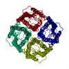
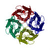
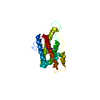
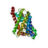
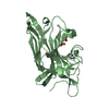
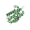
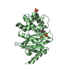
| Entry | Database: PDB / ID: 1h6i | ||||||
|---|---|---|---|---|---|---|---|
| Title | A REFINED STRUCTURE OF HUMAN AQUAPORIN 1 | ||||||
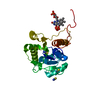
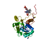
 Components Components | AQUAPORIN-1 | ||||||
 Keywords Keywords | MEMBRANE PROTEIN / WATER CHANNEL / TWO-DIMENSIONAL CRYSTAL | ||||||
| Function / homology |  Function and homology information Function and homology informationnitric oxide transmembrane transporter activity / metanephric descending thin limb development / metanephric proximal straight tubule development / metanephric proximal convoluted tubule segment 2 development / metanephric glomerulus vasculature development / cerebrospinal fluid secretion / lipid digestion / cellular response to salt stress / renal water transport / glycerol transmembrane transporter activity ...nitric oxide transmembrane transporter activity / metanephric descending thin limb development / metanephric proximal straight tubule development / metanephric proximal convoluted tubule segment 2 development / metanephric glomerulus vasculature development / cerebrospinal fluid secretion / lipid digestion / cellular response to salt stress / renal water transport / glycerol transmembrane transporter activity / corticotropin secretion / secretory granule organization / carbon dioxide transmembrane transport / carbon dioxide transmembrane transporter activity / renal water absorption / positive regulation of saliva secretion / Passive transport by Aquaporins / glycerol transmembrane transport / water transmembrane transporter activity / establishment or maintenance of actin cytoskeleton polarity / pancreatic juice secretion / lateral ventricle development / cellular response to mercury ion / potassium ion transmembrane transporter activity / water channel activity / intracellular water homeostasis / cellular response to inorganic substance / intracellularly cGMP-activated cation channel activity / ammonium transmembrane transport / water transport / transepithelial water transport / glomerular filtration / ankyrin-1 complex / ammonium transmembrane transporter activity / camera-type eye morphogenesis / multicellular organismal-level water homeostasis / fibroblast migration / cellular homeostasis / cellular hyperosmotic response / hyperosmotic response / renal water homeostasis / cell volume homeostasis / positive regulation of fibroblast migration / odontogenesis / nitric oxide transport / cGMP-mediated signaling / potassium channel activity / brush border / transmembrane transporter activity / cellular response to nitric oxide / cellular response to retinoic acid / cellular response to cAMP / sensory perception of pain / cellular response to copper ion / ephrin receptor binding / cellular response to dexamethasone stimulus / basal plasma membrane / establishment of localization in cell / brush border membrane / sarcolemma / carbon dioxide transport / negative regulation of cysteine-type endopeptidase activity involved in apoptotic process / wound healing / Erythrocytes take up oxygen and release carbon dioxide / Erythrocytes take up carbon dioxide and release oxygen / potassium ion transport / cellular response to hydrogen peroxide / Vasopressin regulates renal water homeostasis via Aquaporins / cellular response to mechanical stimulus / cellular response to UV / positive regulation of angiogenesis / positive regulation of fibroblast proliferation / apical part of cell / cellular response to hypoxia / basolateral plasma membrane / nuclear membrane / defense response to Gram-negative bacterium / apical plasma membrane / axon / negative regulation of apoptotic process / extracellular exosome / identical protein binding / nucleus / plasma membrane / cytoplasmSimilarity search - Function | ||||||
| Biological species |  HOMO SAPIENS (human) HOMO SAPIENS (human) | ||||||
| Method | ELECTRON CRYSTALLOGRAPHY / electron crystallography / cryo EM / Resolution: 3.54 Å | ||||||
 Authors Authors | De Groot, B.L. / Engel, A. / Grubmuller, H. | ||||||
 Citation Citation | Journal: FEBS Lett / Year: 2001 Title: A refined structure of human aquaporin-1. Authors: B L de Groot / A Engel / H Grubmüller /  Abstract: A refined structure of the human water channel aquaporin-1 is presented. The model rests on the high resolution X-ray structure of the homologous bacterial glycerol transporter GlpF, electron ...A refined structure of the human water channel aquaporin-1 is presented. The model rests on the high resolution X-ray structure of the homologous bacterial glycerol transporter GlpF, electron crystallographic data at 3.8 A resolution and a multiple sequence alignment of the aquaporin superfamily. The crystallographic R and free R values (36.7% and 37.8%) for the refined structure are significantly lower than for previous models. Improved geometry and enhanced stability in molecular dynamics simulations demonstrate a significant improvement of the aquaporin-1 structure. Comparison with previous aquaporin-1 models shows significant differences, not only in the loop regions, but also in the core of the water channel. #1: Journal: J Mol Biol / Year: 2000 Title: The fold of human aquaporin 1. Authors: B L de Groot / J B Heymann / A Engel / K Mitsuoka / Y Fujiyoshi / H Grubmüller / Abstract: The fold of human aquaporin 1 is determined from cryo-electron microscopic data at 4.5 A resolution. The monomeric structure consists of two transmembrane triple helices arranged around a pseudo-2- ...The fold of human aquaporin 1 is determined from cryo-electron microscopic data at 4.5 A resolution. The monomeric structure consists of two transmembrane triple helices arranged around a pseudo-2-fold axis connected by a long flexible extracellular loop. Each triplet contains between its second and third helix a functional loop containing the highly conserved fingerprint NPA motif. These functional loops are assumed to fold inwards between the two triplets, thereby forming the heart of the water channel. The helix topology was determined from the directionality pattern of each of the six transmembrane helices with respect to the membrane, together with constraints defined by the sequence and atomic force microscopy data. The directionality of the helices was determined by collecting the best-fitting orientations resulting from a search through the three-dimensional experimental map for a large number of alpha-helical fragments. Tests on cryo-electron crystallographic bacteriorhodopsin data suggest that our method is generally applicable to determine the topology of helical proteins for which only medium-resolution electron microscopy data are available. | ||||||
| History |
|
- Structure visualization
| Movie |
Movie viewer |
|---|---|
| Structure viewer | Molecule: MolmilJmol/JSmol |
- Downloads & links
-Download
| PDBx/mmCIF format | 1h6i.cif.gz | 45.6 KB | Display | PDBx/mmCIF format |
|---|---|---|---|---|
| PDB format | pdb1h6i.ent.gz | 35.1 KB | Display | PDB format |
| PDBx/mmJSON format | 1h6i.json.gz | Tree view | PDBx/mmJSON format | |
| Others |  Other downloads Other downloads |
-Validation report
| Arichive directory | https://data.pdbj.org/pub/pdb/validation_reports/h6/1h6iftp://data.pdbj.org/pub/pdb/validation_reports/h6/1h6i | HTTPS FTP |
|---|
-Related structure data
| Related structure data | |
|---|---|
| Similar structure data |

-Links
 PDBj
PDBj

- Assembly
| Deposited unit | 
| ||||||||
|---|---|---|---|---|---|---|---|---|---|
| 1 |
| ||||||||
| Unit cell |
|
-Components
| #1: Protein | Mass: 28549.914 Da / Num. of mol.: 1 / Source method: isolated from a natural source / Source: (natural) HOMO SAPIENS (human) / Cell: ERYTHROCYTE / References: UniProt: P29972 |
|---|
-Experimental details
-Experiment
| Experiment | Method: ELECTRON CRYSTALLOGRAPHY |
|---|---|
| EM experiment | Aggregation state: 2D ARRAY / 3D reconstruction method: electron crystallography |
- Sample preparation
| Component | Name: HUMAN AQUAPORIN 1Aquaporin-1 / Type: COMPLEX |
|---|---|
| Specimen | Embedding applied: NO / Shadowing applied: NO / Staining applied: NO / Vitrification applied: YES |
-Data collection
| Microscopy | Model: JEOL 3000SFF |
|---|---|
| Electron gun | Electron source: FIELD EMISSION GUN / Accelerating voltage: 300 kV / Illumination mode: FLOOD BEAM |
| Electron lens | Mode: DIFFRACTION |
| Detector | Date: Jun 7, 1996 |
| Radiation wavelength | Relative weight: 1 |
- Processing
| Software | Name: CNS / Version: 1 / Classification: refinement | ||||||||||||||||||||||||||||||||||||||||||||||||||||||||||||
|---|---|---|---|---|---|---|---|---|---|---|---|---|---|---|---|---|---|---|---|---|---|---|---|---|---|---|---|---|---|---|---|---|---|---|---|---|---|---|---|---|---|---|---|---|---|---|---|---|---|---|---|---|---|---|---|---|---|---|---|---|---|
| 3D reconstruction | Symmetry type: 2D CRYSTAL | ||||||||||||||||||||||||||||||||||||||||||||||||||||||||||||
| Refinement | Starting model: THE MODEL WAS DERIVED USING ELECTRON DIFFRACTION DATA ON 2D CRYSTALS Resolution: 3.54→30.04 Å / Rfactor Rfree error: 0.017 / Isotropic thermal model: GROUP / Cross valid method: THROUGHOUT / σ(F): 0
| ||||||||||||||||||||||||||||||||||||||||||||||||||||||||||||
| Solvent computation | Solvent model: FLAT MODEL / Bsol: 18.5467 Å2 / ksol: 0.09599202 e/Å3 | ||||||||||||||||||||||||||||||||||||||||||||||||||||||||||||
| Displacement parameters | Biso mean: 44.4 Å2
| ||||||||||||||||||||||||||||||||||||||||||||||||||||||||||||
| Refine analyze |
| ||||||||||||||||||||||||||||||||||||||||||||||||||||||||||||
| Refinement step | Cycle: LAST / Resolution: 3.54→30.04 Å
| ||||||||||||||||||||||||||||||||||||||||||||||||||||||||||||
| Refine LS restraints |
| ||||||||||||||||||||||||||||||||||||||||||||||||||||||||||||
| LS refinement shell | Resolution: 3.5→3.72 Å / Rfactor Rfree error: 0.058 / Total num. of bins used: 6
| ||||||||||||||||||||||||||||||||||||||||||||||||||||||||||||
| Xplor file | Serial no: 1 / Param file: PROTEIN_REP.PARAM / Topol file: PROTEIN.TOP |
