Movie
Movie Controller


+ Open data

- Basic information
| Entry | Database: EMDB / ID: EMD-8146 | |||||||||
|---|---|---|---|---|---|---|---|---|---|---|
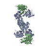
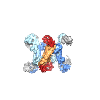
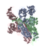
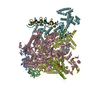
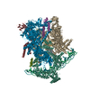
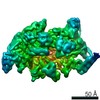
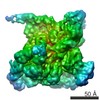
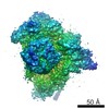
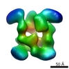
| Title | Methanococcus jannaschii box C/D sRNP | |||||||||
 Map data Map data | None | |||||||||
 Sample Sample |
| |||||||||
| Biological species |    Methanocaldococcus jannaschii (archaea) Methanocaldococcus jannaschii (archaea) | |||||||||
| Method | single particle reconstruction / cryo EM / Resolution: 9.0 Å | |||||||||
 Authors Authors | Yip WSV / Shigematsu H / Taylor DW / Baserga SJ | |||||||||
| Funding support |  United States, 1 items United States, 1 items
| |||||||||
 Citation Citation | Journal: Nucleic Acids Res / Year: 2016 Title: Box C/D sRNA stem ends act as stabilizing anchors for box C/D di-sRNPs. Authors: W S Vincent Yip / Hideki Shigematsu / David W Taylor / Susan J Baserga /  Abstract: Ribosomal RNA (rRNA) modifications are essential for ribosome function in all cellular organisms. Box C/D small (nucleolar) ribonucleoproteins [s(no)RNPs] catalyze 2'-O-methylation, one rRNA ...Ribosomal RNA (rRNA) modifications are essential for ribosome function in all cellular organisms. Box C/D small (nucleolar) ribonucleoproteins [s(no)RNPs] catalyze 2'-O-methylation, one rRNA modification type in Eukarya and Archaea. Negatively stained electron microscopy (EM) models of archaeal box C/D sRNPs have demonstrated the dimeric sRNP (di-sRNP) architecture, which has been corroborated by nuclear magnetic resonance (NMR) studies. Due to limitations of the structural techniques, the orientation of the box C/D sRNAs has remained unclear. Here, we have used cryo-EM to elucidate the sRNA orientation in a M. jannaschii box C/D di-sRNP. The cryo-EM reconstruction suggests a parallel orientation of the two sRNAs. Biochemical and structural analyses of sRNPs assembled with mutant sRNAs indicate a potential interaction between the sRNA stem ends. Our results suggest that the parallel arrangement of the sRNAs juxtaposes their stem ends into close proximity to allow for a stabilizing interaction that helps maintain the di-sRNP architecture. | |||||||||
| History |
|
- Structure visualization
| Movie |
 Movie viewer Movie viewer |
|---|---|
| Structure viewer | EM map: SurfViewMolmilJmol/JSmol |
| Supplemental images |
- Downloads & links
-EMDB archive
| Map data | emd_8146.map.gz | 85.2 MB | EMDB map data format | |
|---|---|---|---|---|
| Header (meta data) | emd-8146-v30.xmlemd-8146.xml | 20.1 KB 20.1 KB | Display Display | EMDB header |
| Images |  emd_8146.png emd_8146.png | 54.8 KB | ||
| Archive directory |  http://ftp.pdbj.org/pub/emdb/structures/EMD-8146ftp://ftp.pdbj.org/pub/emdb/structures/EMD-8146 http://ftp.pdbj.org/pub/emdb/structures/EMD-8146ftp://ftp.pdbj.org/pub/emdb/structures/EMD-8146 | HTTPS FTP |
-Related structure data
| Similar structure data |
|---|

-Links
| EMDB pages | EMDB (EBI/PDBe) / EMDataResource |
|---|
-Map
| File | Download / File: emd_8146.map.gz / Format: CCP4 / Size: 91.1 MB / Type: IMAGE STORED AS FLOATING POINT NUMBER (4 BYTES) | ||||||||||||||||||||||||||||||||||||||||||||||||||||||||||||||||||||
|---|---|---|---|---|---|---|---|---|---|---|---|---|---|---|---|---|---|---|---|---|---|---|---|---|---|---|---|---|---|---|---|---|---|---|---|---|---|---|---|---|---|---|---|---|---|---|---|---|---|---|---|---|---|---|---|---|---|---|---|---|---|---|---|---|---|---|---|---|---|
| Annotation | None | ||||||||||||||||||||||||||||||||||||||||||||||||||||||||||||||||||||
| Projections & slices | Image control
Images are generated by Spider. | ||||||||||||||||||||||||||||||||||||||||||||||||||||||||||||||||||||
| Voxel size | X=Y=Z: 1.247 Å | ||||||||||||||||||||||||||||||||||||||||||||||||||||||||||||||||||||
| Density |
| ||||||||||||||||||||||||||||||||||||||||||||||||||||||||||||||||||||
| Symmetry | Space group: 1 | ||||||||||||||||||||||||||||||||||||||||||||||||||||||||||||||||||||
| Details | EMDB XML:
CCP4 map header:
| ||||||||||||||||||||||||||||||||||||||||||||||||||||||||||||||||||||
 Z (Sec.)
 Y (Row.)
 X (Col.)


-Supplemental data
- Sample components
-Entire : Methanococcus janaschii box C/D sRNP
| Entire | Name: Methanococcus janaschii box C/D sRNP |
|---|---|
| Components |
|
-Supramolecule #1: Methanococcus janaschii box C/D sRNP
| Supramolecule | Name: Methanococcus janaschii box C/D sRNP / type: complex / ID: 1 / Parent: 0 / Macromolecule list: all |
|---|---|
| Source (natural) | Organism: Methanocaldococcus jannaschii (archaea) |
| Molecular weight | Theoretical: 366 KDa |
-Macromolecule #1: L7Ae
| Macromolecule | Name: L7Ae / type: protein_or_peptide / ID: 1 / Enantiomer: LEVO |
|---|---|
| Source (natural) | Organism: Methanocaldococcus jannaschii (archaea) |
| Recombinant expression | Organism:  Escherichia coli (E. coli) Escherichia coli (E. coli) |
| Sequence | String: MAVYVKFKVP EEIQKELLDA VAKAQKIKKG ANEVTKAVER GIAKLVIIAE DVKPEEVVAH LPYLCEEKGI PYAYVASKQD LGKAAGLEVA ASSVAIINEG DAEELKVLIE KVNVLKQ |
-Macromolecule #2: Nop5
| Macromolecule | Name: Nop5 / type: protein_or_peptide / ID: 2 / Enantiomer: LEVO |
|---|---|
| Source (natural) | Organism: Methanocaldococcus jannaschii (archaea) |
| Recombinant expression | Organism: Escherichia coli (E. coli) |
| Sequence | String: MIYVTFTPYG AFGVKDNKEV SGLEDIEYKK LFNEEEIPDI MFKLKTQPNK IADELKEEWG DEIKLETLST EPFNIGEFLR NNLFKVGKEL GYFNNYDEFR KKMHYWSTEL TKKVIKSYAQ QKDKIIIQVA EAISDLDKTL NLLSERLREW YSLYFPELDH LVNKHEVYAN ...String: MIYVTFTPYG AFGVKDNKEV SGLEDIEYKK LFNEEEIPDI MFKLKTQPNK IADELKEEWG DEIKLETLST EPFNIGEFLR NNLFKVGKEL GYFNNYDEFR KKMHYWSTEL TKKVIKSYAQ QKDKIIIQVA EAISDLDKTL NLLSERLREW YSLYFPELDH LVNKHEVYAN LITKLGKRKN FTKSQLKKIL PSKLAGKIAE AAKNSMGGEL EDYDLDVIVK FAEEINHLYE KRKELYNYLE KLMNEEAPNI TKLAGVSLGA RLIGLAGGLE KLAKMPASTI QVLGAEKALF AHLRMGVEPP KHGIIYNHPL IQGSPHWQRG KIARALACKL AIAARADYVG DYIADELLEK LNKRVEEIRR KYPKPPK |
-Macromolecule #3: Fibrillarin
| Macromolecule | Name: Fibrillarin / type: protein_or_peptide / ID: 3 / Enantiomer: LEVO |
|---|---|
| Source (natural) | Organism: Methanocaldococcus jannaschii (archaea) |
| Recombinant expression | Organism: Escherichia coli (E. coli) |
| Sequence | String: MEDIKIKEIF ENIYEVDLGD GLKRIATKSI VKGKKVYDEK IIKIGDEEYR IWNPNKSKLA AAIIKGLKVM PIKRDSKILY LGASAGTTPS HVADIADKGI VYAIEYAPRI MRELLDACAE RENIIPILGD ANKPQEYANI VEKVDVIYED VAQPNQAEIL IKNAKWFLKK ...String: MEDIKIKEIF ENIYEVDLGD GLKRIATKSI VKGKKVYDEK IIKIGDEEYR IWNPNKSKLA AAIIKGLKVM PIKRDSKILY LGASAGTTPS HVADIADKGI VYAIEYAPRI MRELLDACAE RENIIPILGD ANKPQEYANI VEKVDVIYED VAQPNQAEIL IKNAKWFLKK GGYGMIAIKA RSIDVTKDPK EIFKEQKEIL EAGGFKIVDE VDIEPFEKDH VMFVGIWEGK |
-Macromolecule #4: Methanococcus jannaschii sR8 box C/D sRNA
| Macromolecule | Name: Methanococcus jannaschii sR8 box C/D sRNA / type: rna / ID: 4 |
|---|---|
| Sequence | String: AAAUCGCCAA UGAUGACGAU UGGCUUUGCU GAGUCUGUGA UGAACCGUAU GAGCACUGAG GCGAUUU |
-Experimental details
-Structure determination
| Method | cryo EM |
|---|---|
 Processing Processing | single particle reconstruction |
| Aggregation state | particle |
-Sample preparation
| Buffer | pH: 7.5 Component:
| ||||||||||||
|---|---|---|---|---|---|---|---|---|---|---|---|---|---|
| Grid | Model: Electron Microscopy Sciences / Material: COPPER / Mesh: 400 / Support film - Material: CARBON / Support film - topology: CONTINUOUS / Pretreatment - Type: PLASMA CLEANING / Pretreatment - Atmosphere: OTHER | ||||||||||||
| Vitrification | Cryogen name: ETHANE / Chamber humidity: 100 % / Chamber temperature: 281 K / Instrument: FEI VITROBOT MARK III Details: Blotting time 5 seconds; Blot offset -1 mm; plunged into liquid ethane (FEI VITROBOT MARK III).. |
- Electron microscopy
| Microscope | FEI TECNAI F20 |
|---|---|
| Electron beam | Acceleration voltage: 200 kV / Electron source: FIELD EMISSION GUN |
| Electron optics | Illumination mode: FLOOD BEAM / Imaging mode: BRIGHT FIELDBright-field microscopy / Cs: 2.0 mm / Nominal defocus max: 4.0 µm / Nominal defocus min: 2.5 µm / Nominal magnification: 29000 |
| Sample stage | Cooling holder cryogen: NITROGEN |
| Image recording | #0 - Image recording ID: 1 / #0 - Film or detector model: GATAN K2 SUMMIT (4k x 4k) / #0 - Detector mode: COUNTING / #0 - Number grids imaged: 1 / #0 - Number real images: 833 / #0 - Average exposure time: 4.25 sec. / #0 - Average electron dose: 30.0 e/Å2 / #0 - Details: Session 1 / #1 - Image recording ID: 2 / #1 - Film or detector model: GATAN K2 SUMMIT (4k x 4k) / #1 - Detector mode: COUNTING / #1 - Number grids imaged: 1 / #1 - Number real images: 281 / #1 - Average exposure time: 7.25 sec. / #1 - Average electron dose: 30.0 e/Å2 / #1 - Details: Session 2 / #2 - Image recording ID: 3 / #2 - Film or detector model: GATAN K2 SUMMIT (4k x 4k) / #2 - Detector mode: COUNTING / #2 - Number grids imaged: 1 / #2 - Number real images: 546 / #2 - Average exposure time: 6.25 sec. / #2 - Average electron dose: 30.0 e/Å2 / #2 - Details: Session 3 |
| Experimental equipment |  Model: Tecnai F20 / Image courtesy: FEI Company |
-Image processing
| Particle selection | Number selected: 164282 | ||||||
|---|---|---|---|---|---|---|---|
| CTF correction | Software:
Details: CTFFIND3 in Relion was used for CTF estimation | ||||||
| Startup model | Type of model: EMDB MAP EMDB ID: Details: A Methanococcus jannaschii negatively-stained EM map low-passed to 60 Angstrom resolution was used as the initial model for 3D reconstruction. | ||||||
| Initial angle assignment | Type: PROJECTION MATCHING / Software - Name: Reion (ver. 1.3) / Software - details: Relion 3D auto-refine Details: Refinement was performed using Relion 3D auto-refine. Initial angular sampling was 7.5 degrees; initial offset range was 5 pixels; initial offset step was 1 pixel. | ||||||
| Final 3D classification | Number classes: 3 / Avg.num./class: 24000 / Software - Name: RELION (ver. 1.3) / Software - details: Relion 3D Classification Details: 3D classification was performed on "shiny" particles that have beam-induced motions corrected by lm-bfgs alignparts. | ||||||
| Final angle assignment | Type: PROJECTION MATCHING / Software - Name: RELION (ver. 1.3) / Software - details: Relion 3D auto-refine Details: Refinement was performed using Relion 3D auto-refine. Angular sampling, offset range, and offset step sizes were automatically adjusted in Relion. | ||||||
| Final reconstruction | Applied symmetry - Point group: C2 (2 fold cyclic) / Resolution.type: BY AUTHOR / Resolution: 9.0 Å / Resolution method: FSC 0.143 CUT-OFF / Software - Name: RELION (ver. 1.3) / Software - details: 3D auto-refine Details: 3D refinement was performed on "shiny" particles that have beam-induced motions corrected by lm-bfgs alignparts. Number images used: 32771 | ||||||
| Image recording ID | 1 |
-Atomic model buiding 1
| Initial model |
| ||||||||
|---|---|---|---|---|---|---|---|---|---|
| Details | To perform rigid-body docking, two copies of the co-crystal structure from P. furiosus [PDB ID 3NVI, each containing two copies of L7Ae, kink-turn RNA, and Nop5 lacking the NTD (amino acids 127-373)] were docked as rigid bodies into the center of the EM volume (using "Fit in Map" in Chimera) based on prior knowledge of the location of the Nop5 coiled-coil domain. Subsequently, four copies of P. furiosus Nop5 N-terminal domain (amino acids 8-126) and fibrillarin from PDB ID 2NNW were docked into the four corners of the volume using "Fit to Segments" in Chimera without changing the relative orientation between the two proteins. To refine the docking, a simultaneous multi-fragment docking refinement was performed using Collage in Situs 2.8. | ||||||||
| Refinement | Space: REAL / Protocol: RIGID BODY FIT / Target criteria: Correlation |