 Movie
Movie Controller

[English] 日本語
 Yorodumi
- EMDB-6426: Negative-stain electron microscopy of a full-length transmembrane... -
+ Open data

- Basic information
| Entry | Database: EMDB / ID: EMD-6426 | |||||||||
|---|---|---|---|---|---|---|---|---|---|---|
| Title | Negative-stain electron microscopy of a full-length transmembrane receptor tyrosine kinase (PDGFR) in complex with PDGFB | |||||||||
 Map data Map data | Reconstruction of PDGF-B in complex with full-length PDGFR-Beta | |||||||||
 Sample Sample |
| |||||||||
 Keywords Keywords |  receptor tyrosine kinase / membrane protein / cancer / signal transduction / phosphorylation receptor tyrosine kinase / membrane protein / cancer / signal transduction / phosphorylation | |||||||||
| Function / homology |  Function and homology information Function and homology informationmetanephric glomerular mesangial cell development / positive regulation of vascular associated smooth muscle cell dedifferentiation / platelet-derived growth factor complex / positive regulation of metanephric mesenchymal cell migration / negative regulation of phosphatidylinositol biosynthetic process / platelet activating factor receptor activity / platelet-derived growth factor receptor activity / platelet-derived growth factor beta-receptor activity / cell migration involved in coronary angiogenesis / metanephric glomerular mesangial cell proliferation involved in metanephros development ...metanephric glomerular mesangial cell development / positive regulation of vascular associated smooth muscle cell dedifferentiation / platelet-derived growth factor complex / positive regulation of metanephric mesenchymal cell migration / negative regulation of phosphatidylinositol biosynthetic process / platelet activating factor receptor activity / platelet-derived growth factor receptor activity / platelet-derived growth factor beta-receptor activity / cell migration involved in coronary angiogenesis / metanephric glomerular mesangial cell proliferation involved in metanephros development / positive regulation of glomerular filtration / positive regulation of metanephric mesenchymal cell migration by platelet-derived growth factor receptor-beta signaling pathway / smooth muscle cell chemotaxis / metanephric glomerular capillary formation / cellular response to mycophenolic acid / superoxide-generating NADPH oxidase activator activity / negative regulation of vascular associated smooth muscle cell differentiation / cell migration involved in vasculogenesis / protein kinase C signaling / aorta morphogenesis / positive regulation of hyaluronan biosynthetic process / positive regulation of cell proliferation by VEGF-activated platelet derived growth factor receptor signaling pathway / platelet-derived growth factor binding / retina vasculature development in camera-type eye / vascular endothelial growth factor binding / cardiac myofibril assembly / phosphatidylinositol metabolic process / positive regulation of glomerular mesangial cell proliferation / positive regulation of chemotaxis / interleukin-18-mediated signaling pathway / Signaling by PDGF / paracrine signaling / platelet-derived growth factor receptor binding / positive regulation of vascular associated smooth muscle cell migration / positive regulation of cell-substrate adhesion / positive regulation of DNA biosynthetic process / cellular response to platelet-derived growth factor stimulus / positive regulation of smooth muscle cell migration / positive regulation of calcium ion import / chemoattractant activity / platelet-derived growth factor receptor-beta signaling pathway / monocyte chemotaxis / positive regulation of phosphoprotein phosphatase activity / positive regulation of cell division / negative regulation of platelet activation / platelet-derived growth factor receptor signaling pathway / Non-integrin membrane-ECM interactions / embryonic placenta development / positive regulation of blood vessel endothelial cell migration / positive regulation of phospholipase C activity / positive regulation of calcium-mediated signaling / positive regulation of protein autophosphorylation / positive regulation of vascular associated smooth muscle cell proliferation / collagen binding / reactive oxygen species metabolic process / positive regulation of endothelial cell proliferation / lysosomal lumen / cell chemotaxis / Downstream signal transduction / positive regulation of mitotic nuclear division / negative regulation of miRNA transcription / platelet alpha granule lumen / negative regulation of protein binding / regulation of actin cytoskeleton organization / positive regulation of smooth muscle cell proliferation / growth factor activity / positive regulation of MAP kinase activity / receptor protein-tyrosine kinase / cellular response to growth factor stimulus / response to wounding / Golgi lumen / positive regulation of miRNA transcription / peptidyl-tyrosine phosphorylation / cell surface receptor protein tyrosine kinase signaling pathway / Constitutive Signaling by Aberrant PI3K in Cancer / positive regulation of reactive oxygen species metabolic process / positive regulation of fibroblast proliferation / positive regulation of peptidyl-tyrosine phosphorylation / Platelet degranulation / PIP3 activates AKT signaling / gene expression / heart development / PI5P, PP2A and IER3 Regulate PI3K/AKT Signaling / cytoplasmic vesicle / RAF/MAP kinase cascade / basolateral plasma membrane / collagen-containing extracellular matrix / protein tyrosine kinase activity / positive regulation of MAPK cascade / protein autophosphorylation / positive regulation of ERK1 and ERK2 cascade / positive regulation of phosphatidylinositol 3-kinase/protein kinase B signal transduction / receptor complex / protein kinase activity / positive regulation of cell migration / apical plasma membrane / protein heterodimerization activity / endoplasmic reticulum lumen / Golgi membrane / negative regulation of gene expressionSimilarity search - Function | |||||||||
| Biological species |  Homo sapiens (human) Homo sapiens (human) | |||||||||
| Method | single particle reconstruction / negative staining / Resolution: 27.0 Å | |||||||||
 Authors Authors | Chen PH / Unger VM / He X | |||||||||
 Citation Citation | Journal: J Mol Biol / Year: 2015 Title: Structure of Full-Length Human PDGFRβ Bound to Its Activating Ligand PDGF-B as Determined by Negative-Stain Electron Microscopy. Authors: Po-Han Chen / Vinzenz Unger / Xiaolin He /  Abstract: Members of the receptor tyrosine kinases (RTKs) regulate important cellular functions such as cell growth and migration, which are key steps in angiogenesis, in organ morphogenesis and in the ...Members of the receptor tyrosine kinases (RTKs) regulate important cellular functions such as cell growth and migration, which are key steps in angiogenesis, in organ morphogenesis and in the unregulated states, cancer formation. One long-standing puzzle regarding RTKs centers on how the extracellular domain (ECD), which detects and binds to growth factors, is coupled with the intracellular domain kinase activation. While extensive structural works on the soluble portions of RTKs have provided critical insights into RTK structures and functions, lack of a full-length receptor structure has hindered a comprehensive overview of RTK activation. In this study, we successfully purified and determined a 27-Å-resolution structure of PDGFRβ [a full-length human platelet-derived growth factor receptor], in complex with its ligand PDGF-B. In the ligand-stimulated complex, two PDGFRβs assemble into a dimer via an extensive interface essentially running along the full-length of the receptor, suggesting that the membrane-proximal region, the transmembrane helix and the kinase domain of PDGFRβ are involved in dimerization. Major structural differences are seen between the full-length and soluble ECD structures, rationalizing previous experimental data on how membrane-proximal domains modulate receptor ligand-binding affinity and dimerization efficiency. Also, in contrast to the 2-fold symmetry of the ECD, the intracellular kinase domains adopt an asymmetric dimer arrangement, in agreement with prior observations for the closely related KIT receptor. In essence, the structure provides a first glimpse into how platelet-derived growth factor receptor ECD, upon ligand stimulation, is coupled to its intracellular domain kinase activation. | |||||||||
| History |
|
- Structure visualization
| Movie |
Movie viewer |
|---|---|
| Structure viewer | EM map: SurfViewMolmilJmol/JSmol |
| Supplemental images |

- Downloads & links
-EMDB archive
| Map data | emd_6426.map.gz | 536.4 KB | EMDB map data format | |
|---|---|---|---|---|
| Header (meta data) | emd-6426-v30.xmlemd-6426.xml | 11.1 KB 11.1 KB | Display Display | EMDB header |
| Images | 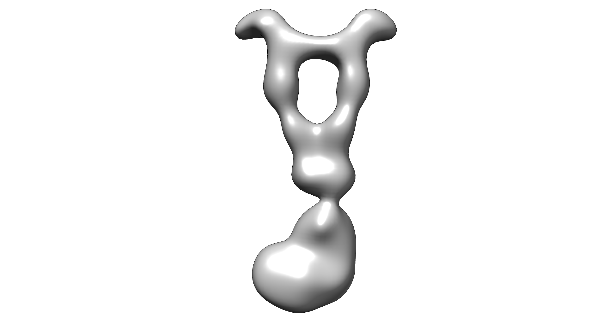 emd_6426.png emd_6426.png | 151.1 KB | ||
| Archive directory |  http://ftp.pdbj.org/pub/emdb/structures/EMD-6426ftp://ftp.pdbj.org/pub/emdb/structures/EMD-6426 http://ftp.pdbj.org/pub/emdb/structures/EMD-6426ftp://ftp.pdbj.org/pub/emdb/structures/EMD-6426 | HTTPS FTP |
-Related structure data
| Similar structure data |
|---|
-Links
| EMDB pages | EMDB (EBI/PDBe) / EMDataResource |
|---|---|
| Related items in Molecule of the Month |


-Map
| File | Download / File: emd_6426.map.gz / Format: CCP4 / Size: 5 MB / Type: IMAGE STORED AS FLOATING POINT NUMBER (4 BYTES) | ||||||||||||||||||||||||||||||||||||||||||||||||||||||||||||||||||||
|---|---|---|---|---|---|---|---|---|---|---|---|---|---|---|---|---|---|---|---|---|---|---|---|---|---|---|---|---|---|---|---|---|---|---|---|---|---|---|---|---|---|---|---|---|---|---|---|---|---|---|---|---|---|---|---|---|---|---|---|---|---|---|---|---|---|---|---|---|---|
| Annotation | Reconstruction of PDGF-B in complex with full-length PDGFR-Beta | ||||||||||||||||||||||||||||||||||||||||||||||||||||||||||||||||||||
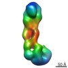
| Voxel size | X=Y=Z: 3.71 Å | ||||||||||||||||||||||||||||||||||||||||||||||||||||||||||||||||||||
| Density |
| ||||||||||||||||||||||||||||||||||||||||||||||||||||||||||||||||||||
| Symmetry | Space group: 1 | ||||||||||||||||||||||||||||||||||||||||||||||||||||||||||||||||||||
| Details | EMDB XML:
CCP4 map header:
| ||||||||||||||||||||||||||||||||||||||||||||||||||||||||||||||||||||
-Supplemental data
- Sample components
-Entire : human PDGF-B bound to PDGFR-Beta
| Entire | Name: human PDGF-B bound to PDGFR-Beta |
|---|---|
| Components |
|
-Supramolecule #1000: human PDGF-B bound to PDGFR-Beta
| Supramolecule | Name: human PDGF-B bound to PDGFR-Beta / type: sample / ID: 1000 Details: The sample is cross-linked via the GraFix method after the gel filtration step. Oligomeric state: a PDGF-B dimer bound to two PDGFR-Beta / Number unique components: 2 |
|---|---|
| Molecular weight | Experimental: 300 KDa / Theoretical: 300 KDa / Method: gel filtration and SDS-PAGE analysis |
-Macromolecule #1: Platelet-derived growth factor receptor beta
| Macromolecule | Name: Platelet-derived growth factor receptor beta / type: protein_or_peptide / ID: 1 / Name.synonym: PDGFR-Beta Details: N-terminal Flag tag is attached after a secretion signal derived from Gaussia luciferase. Number of copies: 2 / Oligomeric state: Dimer / Recombinant expression: Yes |
|---|---|
| Source (natural) | Organism: Homo sapiens (human) / synonym: Human / Location in cell: plasma membrane |
| Molecular weight | Experimental: 150 KDa / Theoretical: 150 KDa |
| Recombinant expression | Organism: Homo sapiens (human) / Recombinant cell: HEK293GnTI- / Recombinant plasmid: BacMam |
| Sequence | UniProtKB: Platelet-derived growth factor receptor beta |
-Macromolecule #2: Platelet-derived growth factor-BB
| Macromolecule | Name: Platelet-derived growth factor-BB / type: protein_or_peptide / ID: 2 / Name.synonym: PDGF-BB / Details: C-terminal residues 191-241 removed / Number of copies: 2 / Oligomeric state: Dimer / Recombinant expression: Yes |
|---|---|
| Source (natural) | Organism: Homo sapiens (human) / synonym: Human / Location in cell: Extracellular |
| Molecular weight | Experimental: 12 KDa / Theoretical: 12 KDa |
| Recombinant expression | Organism: Homo sapiens (human) / Recombinant cell: HEK293GnTI- / Recombinant plasmid: BacMam |
| Sequence | UniProtKB: Platelet-derived growth factor subunit B |
-Experimental details
-Structure determination
| Method | negative staining |
|---|---|
 Processing Processing | single particle reconstruction |
| Aggregation state | particle |
-Sample preparation
| Concentration | 0.02 mg/mL |
|---|---|
| Buffer | pH: 7.6 Details: 150 mM NaCl, 10 mM HEPES, 0.01% LMNG, ~20% glycerol |
| Staining | Type: NEGATIVE Details: Grids with adsorbed protein were floated on 0.75% w/v uranyl formate for 20 seconds. |
| Grid | Details: 400 mesh copper grid with ~2.1 nm thin carbon support, glow-discharged in ambient air atmosphere |
| Vitrification | Cryogen name: NONE / Instrument: OTHER |
- Electron microscopy
| Microscope | OTHER |
|---|---|
| Electron beam | Acceleration voltage: 120 kV / Electron source: FIELD EMISSION GUN |
| Electron optics | Illumination mode: FLOOD BEAM / Imaging mode: BRIGHT FIELDBright-field microscopy / Cs: 3.4 mm / Nominal defocus max: 2.0 µm / Nominal defocus min: 0.5 µm / Nominal magnification: 30000 |
| Sample stage | Specimen holder model: SIDE ENTRY, EUCENTRIC / Tilt angle max: 50 |
| Date | Dec 1, 2014 |
| Image recording | Category: CCD / Film or detector model: GENERIC CCD / Number real images: 100 |
| Tilt angle min | 0 |
-Image processing
| Final reconstruction | Algorithm: OTHER / Resolution.type: BY AUTHOR / Resolution: 27.0 Å / Resolution method: OTHER / Software - Name: XMIPP, RELION / Number images used: 4234 |
|---|---|
| Details | Initial models are reconstructed by RCT. Visual inspection of similar classes were pooled, and one initial model was used as an input model for 3D classification into three classes using RELION. |
