 Movie
Movie Controller

[English] 日本語
 Yorodumi
- EMDB-6330: 3D structure of the decameric assembly of the DNA-injection prote... -
+ Open data

- Basic information
| Entry | Database: EMDB / ID: EMD-6330 | |||||||||
|---|---|---|---|---|---|---|---|---|---|---|
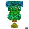
| Title | 3D structure of the decameric assembly of the DNA-injection protein gp12 from bacteriophage Sf6 | |||||||||
 Map data Map data | EM reconstruction of DNA injection protein decameric assembly | |||||||||
 Sample Sample |
| |||||||||
 Keywords Keywords |  bacteriophage / podovirus / DNA injection / decamer / Gram-negative / cell envelope bacteriophage / podovirus / DNA injection / decamer / Gram-negative / cell envelope | |||||||||
| Function / homology | DNA transfer protein / DNA/protein translocase of phage P22 injectosome / Gene 12 protein Function and homology information Function and homology information | |||||||||
| Biological species |  Bacillus phage SF6 (virus) Bacillus phage SF6 (virus) | |||||||||
| Method | single particle reconstruction / negative staining / Resolution: 11.0 Å | |||||||||
 Authors Authors | Zhao H / Speir JA / Matsui T / Lin Z / Liang L / Lynn AY / Varnado B / Weiss TM / Tang L | |||||||||
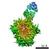
 Citation Citation | Journal: PLoS One / Year: 2016 Title: Structure of a Bacterial Virus DNA-Injection Protein Complex Reveals a Decameric Assembly with a Constricted Molecular Channel. Authors: Haiyan Zhao / Jeffrey A Speir / Tsutomu Matsui / Zihan Lin / Lingfei Liang / Anna Y Lynn / Brittany Varnado / Thomas M Weiss / Liang Tang /  Abstract: The multi-layered cell envelope structure of Gram-negative bacteria represents significant physical and chemical barriers for short-tailed phages to inject phage DNA into the host cytoplasm. Here we ...The multi-layered cell envelope structure of Gram-negative bacteria represents significant physical and chemical barriers for short-tailed phages to inject phage DNA into the host cytoplasm. Here we show that a DNA-injection protein of bacteriophage Sf6, gp12, forms a 465-kDa, decameric assembly in vitro. The electron microscopic structure of the gp12 assembly shows a ~150-Å, mushroom-like architecture consisting of a crown domain and a tube-like domain, which embraces a 25-Å-wide channel that could precisely accommodate dsDNA. The constricted channel suggests that gp12 mediates rapid, uni-directional injection of phage DNA into host cells by providing a molecular conduit for DNA translocation. The assembly exhibits a 10-fold symmetry, which may be a common feature among DNA-injection proteins of P22-like phages and may suggest a symmetry mismatch with respect to the 6-fold symmetric phage tail. The gp12 monomer is highly flexible in solution, supporting a mechanism for translocation of the protein through the conduit of the phage tail toward the host cell envelope, where it assembles into a DNA-injection device. | |||||||||
| History |
|
- Structure visualization
| Movie |
Movie viewer |
|---|---|
| Structure viewer | EM map: SurfViewMolmilJmol/JSmol |
| Supplemental images |

- Downloads & links
-EMDB archive
| Map data | emd_6330.map.gz | 5.5 MB | EMDB map data format | |
|---|---|---|---|---|
| Header (meta data) | emd-6330-v30.xmlemd-6330.xml | 10.9 KB 10.9 KB | Display Display | EMDB header |
| FSC (resolution estimation) | emd_6330_fsc.xml | 4.4 KB | Display | FSC data file |
| Images | 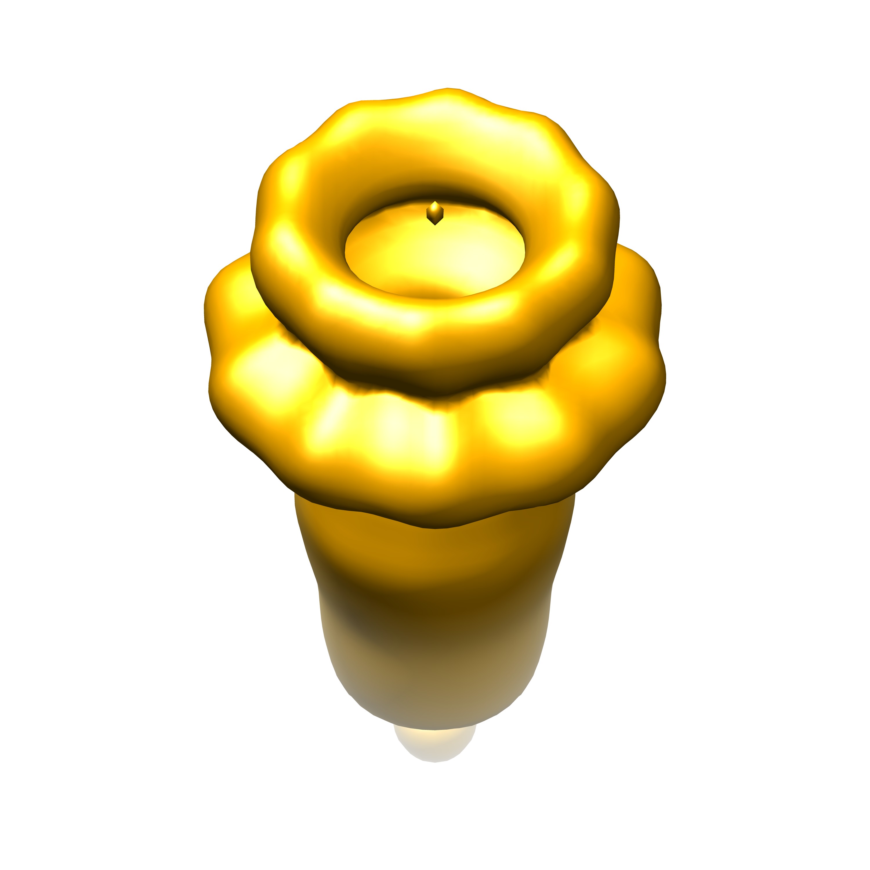 emd_6330.jpg emd_6330.jpg | 298.2 KB | ||
| Archive directory |  http://ftp.pdbj.org/pub/emdb/structures/EMD-6330ftp://ftp.pdbj.org/pub/emdb/structures/EMD-6330 http://ftp.pdbj.org/pub/emdb/structures/EMD-6330ftp://ftp.pdbj.org/pub/emdb/structures/EMD-6330 | HTTPS FTP |
-Related structure data
| Similar structure data |
|---|

-Links
| EMDB pages | EMDB (EBI/PDBe) / EMDataResource |
|---|
-Map
| File | Download / File: emd_6330.map.gz / Format: CCP4 / Size: 7.8 MB / Type: IMAGE STORED AS FLOATING POINT NUMBER (4 BYTES) | ||||||||||||||||||||||||||||||||||||||||||||||||||||||||||||||||||||
|---|---|---|---|---|---|---|---|---|---|---|---|---|---|---|---|---|---|---|---|---|---|---|---|---|---|---|---|---|---|---|---|---|---|---|---|---|---|---|---|---|---|---|---|---|---|---|---|---|---|---|---|---|---|---|---|---|---|---|---|---|---|---|---|---|---|---|---|---|---|
| Annotation | EM reconstruction of DNA injection protein decameric assembly | ||||||||||||||||||||||||||||||||||||||||||||||||||||||||||||||||||||
| Voxel size | X=Y=Z: 2.73 Å | ||||||||||||||||||||||||||||||||||||||||||||||||||||||||||||||||||||
| Density |
| ||||||||||||||||||||||||||||||||||||||||||||||||||||||||||||||||||||
| Symmetry | Space group: 1 | ||||||||||||||||||||||||||||||||||||||||||||||||||||||||||||||||||||
| Details | EMDB XML:
CCP4 map header:
| ||||||||||||||||||||||||||||||||||||||||||||||||||||||||||||||||||||
-Supplemental data
- Sample components
-Entire : gp12 of bacteriophage Sf6
| Entire | Name: gp12 of bacteriophage Sf6 |
|---|---|
| Components |
|
-Supramolecule #1000: gp12 of bacteriophage Sf6
| Supramolecule | Name: gp12 of bacteriophage Sf6 / type: sample / ID: 1000 / Details: The sample was mostly monodisperse. / Oligomeric state: decamer / Number unique components: 1 |
|---|---|
| Molecular weight | Experimental: 465 KDa / Theoretical: 465 KDa / Method: SDS PAGE and size exclusion |
-Macromolecule #1: product of gene 12
| Macromolecule | Name: product of gene 12 / type: protein_or_peptide / ID: 1 / Name.synonym: gp12 / Number of copies: 10 / Oligomeric state: decamer / Recombinant expression: Yes |
|---|---|
| Source (natural) | Organism: Bacillus phage SF6 (virus) / synonym: phage Sf6 |
| Molecular weight | Experimental: 46.5 KDa / Theoretical: 46.5 KDa |
| Recombinant expression | Organism:  Escherichia coli (E. coli) / Recombinant strain: BL21(DE3) / Recombinant plasmid: pET28b Escherichia coli (E. coli) / Recombinant strain: BL21(DE3) / Recombinant plasmid: pET28b |
| Sequence | UniProtKB: Gene 12 protein |
-Experimental details
-Structure determination
| Method | negative staining |
|---|---|
 Processing Processing | single particle reconstruction |
| Aggregation state | particle |
-Sample preparation
| Concentration | 0.0415 mg/mL |
|---|---|
| Buffer | pH: 7.4 / Details: 20 mM NaPO4, 100 mM NaCl, 1 mM EDTA |
| Staining | Type: NEGATIVE Details: 3 uL sample was applied onto a grid that was plasma cleaned with Ar/O2 mixture (20 sec, full power) |
| Grid | Details: copper 400 mesh with carbon over vinyl |
| Vitrification | Cryogen name: NONE / Instrument: OTHER |
- Electron microscopy
| Microscope | FEI TECNAI F20 |
|---|---|
| Electron beam | Acceleration voltage: 200 kV / Electron source: FIELD EMISSION GUN |
| Electron optics | Illumination mode: FLOOD BEAM / Imaging mode: BRIGHT FIELDBright-field microscopy / Nominal defocus max: -2.45 µm / Nominal defocus min: -0.29 µm / Nominal magnification: 62000 |
| Sample stage | Specimen holder model: GATAN LIQUID NITROGEN |
| Temperature | Average: 90 K |
| Date | Aug 21, 2014 |
| Image recording | Category: CCD / Film or detector model: TVIPS TEMCAM-F416 (4k x 4k) / Number real images: 494 / Average electron dose: 38.89 e/Å2 / Bits/pixel: 8 |
| Experimental equipment |  Model: Tecnai F20 / Image courtesy: FEI Company |
-Image processing
| CTF correction | Details: CTFFIND |
|---|---|
| Final two d classification | Number classes: 23 |
| Final angle assignment | Details: RELION: theta 180 degrees, phi 36 degrees |
| Final reconstruction | Algorithm: OTHER / Resolution.type: BY AUTHOR / Resolution: 11.0 Å / Resolution method: OTHER / Software - Name: RELION / Number images used: 54104 |
| Details | A total of 86783 particles were boxed with a dimension of 128x128 pixels and a pixel size of 2.73 A using the program DoGPicker in the Appion pipeline. Image analysis and 3D reconstruction were performed using RELION. No symmetry was applied during 2D classification. After 2D classification, 54104 particles associated with high-quality class averages were included for the subsequent 3D reconstruction. In the 3D reconstruction, 10-fold rotational symmetry was applied. |
| FSC plot (resolution estimation) | 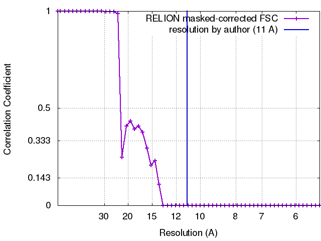 |
