 Movie
Movie Controller

[English] 日本語
 Yorodumi
- EMDB-6329: Electron cryo-microscopy of the 73S Neurospora crassa mitochondri... -
+ Open data

- Basic information
| Entry | Database: EMDB / ID: EMD-6329 | |||||||||
|---|---|---|---|---|---|---|---|---|---|---|
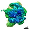
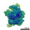
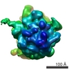
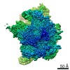
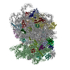
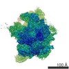
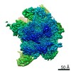
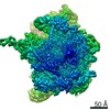
| Title | Electron cryo-microscopy of the 73S Neurospora crassa mitochondrial ribosome | |||||||||
 Map data Map data | 73S mitochondrial ribosome | |||||||||
 Sample Sample |
| |||||||||
 Keywords Keywords |  Mitochondrial ribosome Mitochondrial ribosome | |||||||||
| Biological species |  Neurospora crassa (fungus) Neurospora crassa (fungus) | |||||||||
| Method | single particle reconstruction / cryo EM / Resolution: 7.5 Å | |||||||||
 Authors Authors | Van der Sluis EO / Bauerschmitt H / Becker T / Mielke T / Frauenfeld J / Berninghausen O / Neupert W / Herrmann JM / Beckmann R | |||||||||
 Citation Citation | Journal: Genome Biol Evol / Year: 2015 Title: Parallel Structural Evolution of Mitochondrial Ribosomes and OXPHOS Complexes. Authors: Eli O van der Sluis / Heike Bauerschmitt / Thomas Becker / Thorsten Mielke / Jens Frauenfeld / Otto Berninghausen / Walter Neupert / Johannes M Herrmann / Roland Beckmann /   Abstract: The five macromolecular complexes that jointly mediate oxidative phosphorylation (OXPHOS) in mitochondria consist of many more subunits than those of bacteria, yet, it remains unclear by which ...The five macromolecular complexes that jointly mediate oxidative phosphorylation (OXPHOS) in mitochondria consist of many more subunits than those of bacteria, yet, it remains unclear by which evolutionary mechanism(s) these novel subunits were recruited. Even less well understood is the structural evolution of mitochondrial ribosomes (mitoribosomes): while it was long thought that their exceptionally high protein content would physically compensate for their uniquely low amount of ribosomal RNA (rRNA), this hypothesis has been refuted by structural studies. Here, we present a cryo-electron microscopy structure of the 73S mitoribosome from Neurospora crassa, together with genomic and proteomic analyses of mitoribosome composition across the eukaryotic domain. Surprisingly, our findings reveal that both structurally and compositionally, mitoribosomes have evolved very similarly to mitochondrial OXPHOS complexes via two distinct phases: A constructive phase that mainly acted early in eukaryote evolution, resulting in the recruitment of altogether approximately 75 novel subunits, and a reductive phase that acted during metazoan evolution, resulting in gradual length-reduction of mitochondrially encoded rRNAs and OXPHOS proteins. Both phases can be well explained by the accumulation of (slightly) deleterious mutations and deletions, respectively, in mitochondrially encoded rRNAs and OXPHOS proteins. We argue that the main role of the newly recruited (nuclear encoded) ribosomal- and OXPHOS proteins is to provide structural compensation to the mutationally destabilized mitochondrially encoded components. While the newly recruited proteins probably provide a selective advantage owing to their compensatory nature, and while their presence may have opened evolutionary pathways toward novel mitochondrion-specific functions, we emphasize that the initial events that resulted in their recruitment was nonadaptive in nature. Our framework is supported by population genetic studies, and it can explain the complete structural evolution of mitochondrial ribosomes and OXPHOS complexes, as well as many observed functions of individual proteins. | |||||||||
| History |
|
- Structure visualization
| Movie |
 Movie viewer Movie viewer |
|---|---|
| Structure viewer | EM map: SurfViewMolmilJmol/JSmol |
| Supplemental images |
- Downloads & links
-EMDB archive
| Map data | emd_6329.map.gz | 178.1 MB | EMDB map data format | |
|---|---|---|---|---|
| Header (meta data) | emd-6329-v30.xmlemd-6329.xml | 9.6 KB 9.6 KB | Display Display | EMDB header |
| Images |  400_6329.gif 400_6329.gif 80_6329.gif 80_6329.gif | 72.7 KB 3.8 KB | ||
| Archive directory |  http://ftp.pdbj.org/pub/emdb/structures/EMD-6329ftp://ftp.pdbj.org/pub/emdb/structures/EMD-6329 http://ftp.pdbj.org/pub/emdb/structures/EMD-6329ftp://ftp.pdbj.org/pub/emdb/structures/EMD-6329 | HTTPS FTP |
-Related structure data
| Similar structure data |
|---|


-Links
| EMDB pages | EMDB (EBI/PDBe) / EMDataResource |
|---|---|
| Related items in Molecule of the Month |

-Map
| File | Download / File: emd_6329.map.gz / Format: CCP4 / Size: 185.7 MB / Type: IMAGE STORED AS FLOATING POINT NUMBER (4 BYTES) | ||||||||||||||||||||||||||||||||||||||||||||||||||||||||||||
|---|---|---|---|---|---|---|---|---|---|---|---|---|---|---|---|---|---|---|---|---|---|---|---|---|---|---|---|---|---|---|---|---|---|---|---|---|---|---|---|---|---|---|---|---|---|---|---|---|---|---|---|---|---|---|---|---|---|---|---|---|---|
| Annotation | 73S mitochondrial ribosome | ||||||||||||||||||||||||||||||||||||||||||||||||||||||||||||
| Voxel size | X=Y=Z: 1.23 Å | ||||||||||||||||||||||||||||||||||||||||||||||||||||||||||||
| Density |
| ||||||||||||||||||||||||||||||||||||||||||||||||||||||||||||
| Symmetry | Space group: 1 | ||||||||||||||||||||||||||||||||||||||||||||||||||||||||||||
| Details | EMDB XML:
CCP4 map header:
| ||||||||||||||||||||||||||||||||||||||||||||||||||||||||||||
-Supplemental data
- Sample components
-Entire : 73S mitochondrial ribosome from Neurospora crassa
| Entire | Name: 73S mitochondrial ribosome from Neurospora crassa |
|---|---|
| Components |
|
-Supramolecule #1000: 73S mitochondrial ribosome from Neurospora crassa
| Supramolecule | Name: 73S mitochondrial ribosome from Neurospora crassa / type: sample / ID: 1000 / Number unique components: 1 |
|---|---|
| Molecular weight | Theoretical: 3.7 MDa |
-Supramolecule #1: 73S mitochondrial ribosome
| Supramolecule | Name: 73S mitochondrial ribosome / type: complex / ID: 1 / Name.synonym: 73S mitoribosome / Recombinant expression: No / Database: NCBI / Ribosome-details: ribosome-eukaryote: mitochondrial ALL |
|---|---|
| Source (natural) | Organism: Neurospora crassa (fungus) / Strain: K5-15-23-1 / Organelle: mitochondrion |
| Molecular weight | Theoretical: 3.7 MDa |
-Experimental details
-Structure determination
| Method | cryo EM |
|---|---|
 Processing Processing | single particle reconstruction |
| Aggregation state | particle |
-Sample preparation
| Concentration | 0.3 mg/mL |
|---|---|
| Buffer | pH: 7.5 Details: 30 mM Tris-HCl, pH 7.5, 100 mM NH4Cl, 10 mM MgCl2, 4 mM 2-mercaptoethanol, 0.05% beta-D dodecyl maltoside, 0.0075% cardiolipin |
| Grid | Details: Quantifoil grids pre-coated with 2 nm carbon on top |
| Vitrification | Cryogen name: ETHANE / Chamber humidity: 95 % / Chamber temperature: 95 K / Instrument: FEI VITROBOT MARK IV / Method: Blot for 3 seconds before plunging |
- Electron microscopy
| Microscope | FEI POLARA 300 |
|---|---|
| Electron beam | Acceleration voltage: 300 kV / Electron source: FIELD EMISSION GUN |
| Electron optics | Calibrated magnification: 38900 / Illumination mode: FLOOD BEAM / Imaging mode: BRIGHT FIELDBright-field microscopy / Cs: 2.26 mm / Nominal defocus max: 3.7 µm / Nominal defocus min: 1.4 µm / Nominal magnification: 39000 |
| Sample stage | Specimen holder: nitrogen-cooled / Specimen holder model: SIDE ENTRY, EUCENTRIC |
| Date | Apr 10, 2008 |
| Image recording | Category: FILM / Film or detector model: KODAK SO-163 FILM / Digitization - Scanner: PRIMESCAN / Number real images: 132 / Average electron dose: 20 e/Å2 / Bits/pixel: 16 |
| Experimental equipment |  Model: Tecnai Polara / Image courtesy: FEI Company |
-Image processing
| CTF correction | Details: Micrograph |
|---|---|
| Final angle assignment | Details: Spider |
| Final reconstruction | Algorithm: OTHER / Resolution.type: BY AUTHOR / Resolution: 7.5 Å / Resolution method: OTHER / Software - Name: Spider / Number images used: 208737 |
| Details | Particles were selected using SIGNATURE and processed with SPIDER. |
