 Movie
Movie Controller

[English] 日本語
 Yorodumi
- EMDB-6305: The cryo-EM structure of Meiothermus taiwanensis Lon protease wit... -
+ Open data

- Basic information
| Entry | Database: EMDB / ID: EMD-6305 | |||||||||
|---|---|---|---|---|---|---|---|---|---|---|
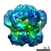
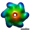
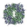
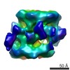
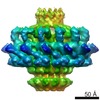
| Title | The cryo-EM structure of Meiothermus taiwanensis Lon protease with ATP and Mg2+ | |||||||||
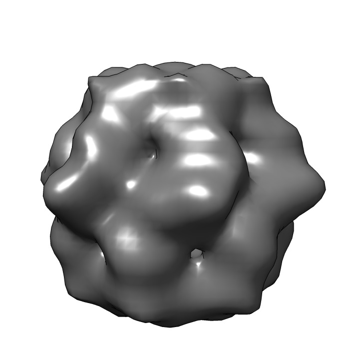
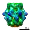
 Map data Map data | Reconstruction of Meiothermus taiwanensis LonA protease with Mg2+ and ATP | |||||||||
 Sample Sample |
| |||||||||
 Keywords Keywords | ATP-dependent proteases | |||||||||
| Biological species |  Meiothermus taiwanensis (bacteria) Meiothermus taiwanensis (bacteria) | |||||||||
| Method |  single particle reconstruction / cryo EM / negative staining / Resolution: 11.8 Å single particle reconstruction / cryo EM / negative staining / Resolution: 11.8 Å | |||||||||
 Authors Authors | Su SC / Chang YC / Chang CI | |||||||||
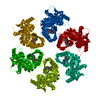
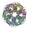
 Citation Citation | Journal: Structure / Year: 2016 Title: Structural Basis for the Magnesium-Dependent Activation and Hexamerization of the Lon AAA+ Protease. Authors: Shih-Chieh Su / Chien-Chu Lin / Hui-Chung Tai / Mu-Yueh Chang / Meng-Ru Ho / C Satheesan Babu / Jiahn-Haur Liao / Shih-Hsiung Wu / Yuan-Chih Chang / Carmay Lim / Chung-I Chang /  Abstract: The Lon AAA+ protease (LonA) plays important roles in protein homeostasis and regulation of diverse biological processes. LonA behaves as a homomeric hexamer in the presence of magnesium (Mg(2+)) and ...The Lon AAA+ protease (LonA) plays important roles in protein homeostasis and regulation of diverse biological processes. LonA behaves as a homomeric hexamer in the presence of magnesium (Mg(2+)) and performs ATP-dependent proteolysis. However, it is also found that LonA can carry out Mg(2+)-dependent degradation of unfolded protein substrate in an ATP-independent manner. Here we show that in the presence of Mg(2+) LonA forms a non-secluded hexameric barrel with prominent openings, which explains why Mg(2+)-activated LonA can operate as a diffusion-based chambered protease to degrade unstructured protein and peptide substrates efficiently in the absence of ATP. A 1.85 Å crystal structure of Mg(2+)-activated protease domain reveals Mg(2+)-dependent remodeling of a substrate-binding loop and a potential metal-binding site near the Ser-Lys catalytic dyad, supported by biophysical binding assays and molecular dynamics simulations. Together, these findings reveal the specific roles of Mg(2+) in the molecular assembly and activation of LonA. | |||||||||
| History |
|
- Structure visualization
| Movie |
 Movie viewer Movie viewer |
|---|---|
| Structure viewer | EM map: SurfViewMolmilJmol/JSmol |
| Supplemental images |
- Downloads & links
-EMDB archive
| Map data | emd_6305.map.gz | 6.6 MB | EMDB map data format | |
|---|---|---|---|---|
| Header (meta data) | emd-6305-v30.xmlemd-6305.xml | 10.5 KB 10.5 KB | Display Display | EMDB header |
| Images | emd_6305.tiff | 163 KB | ||
| Archive directory |  http://ftp.pdbj.org/pub/emdb/structures/EMD-6305ftp://ftp.pdbj.org/pub/emdb/structures/EMD-6305 http://ftp.pdbj.org/pub/emdb/structures/EMD-6305ftp://ftp.pdbj.org/pub/emdb/structures/EMD-6305 | HTTPS FTP |
-Related structure data
-Links
| EMDB pages | EMDB (EBI/PDBe) / EMDataResource |
|---|
-Map
| File | Download / File: emd_6305.map.gz / Format: CCP4 / Size: 7.8 MB / Type: IMAGE STORED AS FLOATING POINT NUMBER (4 BYTES) | ||||||||||||||||||||||||||||||||||||||||||||||||||||||||||||||||||||
|---|---|---|---|---|---|---|---|---|---|---|---|---|---|---|---|---|---|---|---|---|---|---|---|---|---|---|---|---|---|---|---|---|---|---|---|---|---|---|---|---|---|---|---|---|---|---|---|---|---|---|---|---|---|---|---|---|---|---|---|---|---|---|---|---|---|---|---|---|---|
| Annotation | Reconstruction of Meiothermus taiwanensis LonA protease with Mg2+ and ATP | ||||||||||||||||||||||||||||||||||||||||||||||||||||||||||||||||||||
| Voxel size | X=Y=Z: 2.5 Å | ||||||||||||||||||||||||||||||||||||||||||||||||||||||||||||||||||||
| Density |
| ||||||||||||||||||||||||||||||||||||||||||||||||||||||||||||||||||||
| Symmetry | Space group: 1 | ||||||||||||||||||||||||||||||||||||||||||||||||||||||||||||||||||||
| Details | EMDB XML:
CCP4 map header:
| ||||||||||||||||||||||||||||||||||||||||||||||||||||||||||||||||||||
-Supplemental data
- Sample components
-Entire : Meiothermus taiwanensis LonA protease
| Entire | Name: Meiothermus taiwanensis LonA protease |
|---|---|
| Components |
|
-Supramolecule #1000: Meiothermus taiwanensis LonA protease
| Supramolecule | Name: Meiothermus taiwanensis LonA protease / type: sample / ID: 1000 Oligomeric state: One homotetramer of MtaLonA in closed form Number unique components: 1 |
|---|---|
| Molecular weight | Experimental: 540 KDa / Theoretical: 540 KDa / Method: Sedimentation |
-Macromolecule #1: Meiothermus taiwanensis LonA protease
| Macromolecule | Name: Meiothermus taiwanensis LonA protease / type: protein_or_peptide / ID: 1 / Name.synonym: MtaLonA / Oligomeric state: Hexamer / Recombinant expression: Yes |
|---|---|
| Source (natural) | Organism: Meiothermus taiwanensis (bacteria) / synonym: Meiothermus taiwanensis LonA protease |
| Molecular weight | Experimental: 540 KDa / Theoretical: 540 KDa |
| Recombinant expression | Organism: Escherichia coli (E. coli) / Recombinant plasmid: pET21a |
-Experimental details
-Structure determination
| Method | negative staining, cryo EM |
|---|---|
 Processing Processing | single particle reconstruction |
| Aggregation state | particle |
-Sample preparation
| Concentration | 0.12 mg/mL |
|---|---|
| Buffer | pH: 8 Details: 20mM Tris-HCl, 100mM NaCl, 1mM DTT, 10mM MgCl2, 2.5mM ATP |
| Staining | Type: NEGATIVE Details: Grids with adsorbed protein floated on 1% w/v uranyl acetate for 60 seconds. |
| Grid | Details: 200 mesh gold grid with thin carbon support, glow discharged in amylamine atmosphere |
| Vitrification | Cryogen name: ETHANE / Chamber humidity: 100 % / Instrument: FEI VITROBOT MARK IV Method: 1. wait for 10 seconds before blot 2. Blot for 3 seconds before plunging |
- Electron microscopy
| Microscope | FEI TECNAI F20 |
|---|---|
| Electron beam | Acceleration voltage: 200 kV / Electron source: FIELD EMISSION GUN |
| Electron optics | Calibrated magnification: 85600 / Illumination mode: FLOOD BEAM / Imaging mode: BRIGHT FIELDBright-field microscopy / Cs: 2 mm / Nominal defocus max: 3.7 µm / Nominal defocus min: 1.45 µm / Nominal magnification: 62000 |
| Sample stage | Specimen holder model: GATAN LIQUID NITROGEN |
| Alignment procedure | Legacy - Astigmatism: Objective lens astigmatism was corrected at 100,000 times magnification |
| Date | Mar 3, 2014 |
| Image recording | Category: CCD / Film or detector model: GATAN ULTRASCAN 4000 (4k x 4k) / Number real images: 220 / Average electron dose: 20 e/Å2 |
| Experimental equipment |  Model: Tecnai F20 / Image courtesy: FEI Company |
-Image processing
| CTF correction | Details: Each particle |
|---|---|
| Final two d classification | Number classes: 80 |
| Final reconstruction | Resolution.type: BY AUTHOR / Resolution: 11.8 Å / Resolution method: OTHER / Software - Name: EMAN2 / Number images used: 30249 |
| Details | The particle images were interactively selected using EMAN Boxer program |
