 Movie
Movie Controller

[English] 日本語
 Yorodumi
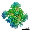
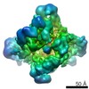
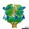
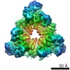
- EMDB-6253: Negative stain reconstruction of the Pex1/Pex6 complex in presenc... -
+ Open data

- Basic information
| Entry | Database: EMDB / ID: EMD-6253 | |||||||||
|---|---|---|---|---|---|---|---|---|---|---|
| Title | Negative stain reconstruction of the Pex1/Pex6 complex in presence of ADP | |||||||||
 Map data Map data | Negative stain reconstruction of the Pex1/Pex6 complex in presence of 3 mM ADP | |||||||||
 Sample Sample |
| |||||||||
 Keywords Keywords | Peroxisome / AAA+ ATPase / Pex1 / Pex6 | |||||||||
| Function / homology |  Function and homology information Function and homology informationATP hydrolysis activity => GO:0016887 / : / protein targeting to peroxisome / protein import into peroxisome matrix, receptor recycling / protein binding / protein import into peroxisome matrix / peroxisome organization / protein transporter activity / peroxisomal membrane / ATPase complex ...ATP hydrolysis activity => GO:0016887 / : / protein targeting to peroxisome / protein import into peroxisome matrix, receptor recycling / protein binding / protein import into peroxisome matrix / peroxisome organization / protein transporter activity / peroxisomal membrane / ATPase complex / protein unfolding / Hydrolases; Acting on acid anhydrides; Acting on acid anhydrides to facilitate cellular and subcellular movement / peroxisome / protein heterodimerization activity / nucleotide binding / ATP hydrolysis activity / ATP binding / membrane / cytoplasm / cytosol Similarity search - Function | |||||||||
| Biological species |  | |||||||||
| Method | single particle reconstruction / negative staining / Resolution: 17.26 Å | |||||||||
 Authors Authors | Gardner BM / Chowdhury S / Lander GC / Martin A | |||||||||
 Citation Citation | Journal: J Mol Biol / Year: 2015 Title: The Pex1/Pex6 complex is a heterohexameric AAA+ motor with alternating and highly coordinated subunits. Authors: Brooke M Gardner / Saikat Chowdhury / Gabriel C Lander / Andreas Martin /  Abstract: Pex1 and Pex6 are Type-2 AAA+ ATPases required for the de novo biogenesis of peroxisomes. Mutations in Pex1 and Pex6 account for the majority of the most severe forms of peroxisome biogenesis ...Pex1 and Pex6 are Type-2 AAA+ ATPases required for the de novo biogenesis of peroxisomes. Mutations in Pex1 and Pex6 account for the majority of the most severe forms of peroxisome biogenesis disorders in humans. Here, we show that the ATP-dependent complex of Pex1 and Pex6 from Saccharomyces cerevisiae is a heterohexamer with alternating subunits. Within the Pex1/Pex6 complex, only the D2 ATPase ring hydrolyzes ATP, while nucleotide binding in the D1 ring promotes complex assembly. ATP hydrolysis by Pex1 is highly coordinated with that of Pex6. Furthermore, Pex15, the membrane anchor required for Pex1/Pex6 recruitment to peroxisomes, inhibits the ATP-hydrolysis activity of Pex1/Pex6. | |||||||||
| History |
|
- Structure visualization
| Movie |
Movie viewer |
|---|---|
| Structure viewer | EM map: SurfViewMolmilJmol/JSmol |
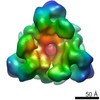
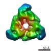
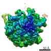
| Supplemental images |
 UCSF Chimera
- Downloads & links
-EMDB archive
| Map data | emd_6253.map.gz | 1.8 MB | EMDB map data format | |
|---|---|---|---|---|
| Header (meta data) | emd-6253-v30.xmlemd-6253.xml | 16.8 KB 16.8 KB | Display Display | EMDB header |
| FSC (resolution estimation) | emd_6253_fsc.xml | 3.4 KB | Display | FSC data file |
| Images | 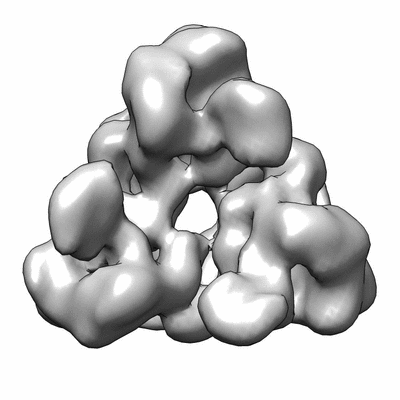 400_6253.gif 400_6253.gif 80_6253.gif 80_6253.gif | 30.2 KB 3.8 KB | ||
| Archive directory |  http://ftp.pdbj.org/pub/emdb/structures/EMD-6253ftp://ftp.pdbj.org/pub/emdb/structures/EMD-6253 http://ftp.pdbj.org/pub/emdb/structures/EMD-6253ftp://ftp.pdbj.org/pub/emdb/structures/EMD-6253 | HTTPS FTP |
-Validation report
| Summary document | emd_6253_validation.pdf.gz | 77.9 KB | Display | EMDB validaton report |
|---|---|---|---|---|
| Full document | emd_6253_full_validation.pdf.gz | 76.9 KB | Display | |
| Data in XML | emd_6253_validation.xml.gz | 494 B | Display | |
| Arichive directory | https://ftp.pdbj.org/pub/emdb/validation_reports/EMD-6253ftp://ftp.pdbj.org/pub/emdb/validation_reports/EMD-6253 | HTTPS FTP |
-Related structure data


-Links
| EMDB pages | EMDB (EBI/PDBe) / EMDataResource |
|---|---|
| Related items in Molecule of the Month |


-Map
| File | Download / File: emd_6253.map.gz / Format: CCP4 / Size: 1.9 MB / Type: IMAGE STORED AS FLOATING POINT NUMBER (4 BYTES) | ||||||||||||||||||||||||||||||||||||||||||||||||||||||||||||||||||||
|---|---|---|---|---|---|---|---|---|---|---|---|---|---|---|---|---|---|---|---|---|---|---|---|---|---|---|---|---|---|---|---|---|---|---|---|---|---|---|---|---|---|---|---|---|---|---|---|---|---|---|---|---|---|---|---|---|---|---|---|---|---|---|---|---|---|---|---|---|---|
| Annotation | Negative stain reconstruction of the Pex1/Pex6 complex in presence of 3 mM ADP | ||||||||||||||||||||||||||||||||||||||||||||||||||||||||||||||||||||
| Voxel size | X=Y=Z: 4.1 Å | ||||||||||||||||||||||||||||||||||||||||||||||||||||||||||||||||||||
| Density |
| ||||||||||||||||||||||||||||||||||||||||||||||||||||||||||||||||||||
| Symmetry | Space group: 1 | ||||||||||||||||||||||||||||||||||||||||||||||||||||||||||||||||||||
| Details | EMDB XML:
CCP4 map header:
| ||||||||||||||||||||||||||||||||||||||||||||||||||||||||||||||||||||
-Supplemental data
- Sample components
-Entire : Pex1/Pex6 complex in presence of 3 mM ADP
| Entire | Name: Pex1/Pex6 complex in presence of 3 mM ADP |
|---|---|
| Components |
|
-Supramolecule #1000: Pex1/Pex6 complex in presence of 3 mM ADP
| Supramolecule | Name: Pex1/Pex6 complex in presence of 3 mM ADP / type: sample / ID: 1000 Details: 22 nM Pex1/Pex6 complex in presence of 3 mM ADP was used for negative stain electron microscopy. The sample was monodisperse. Oligomeric state: Heterohexamer containing three copies of Pex1 and three copies of Pex6 Number unique components: 2 |
|---|---|
| Molecular weight | Theoretical: 707.076 KDa |
-Macromolecule #1: Peroxisomal ATPase PEX1
| Macromolecule | Name: Peroxisomal ATPase PEX1 / type: protein_or_peptide / ID: 1 Name.synonym: Peroxin-1, Peroxisomal assembly protein 1, Peroxisome biogenesis protein PAS1 Number of copies: 3 / Oligomeric state: Heterohexamer / Recombinant expression: Yes |
|---|---|
| Source (natural) | Organism: |
| Molecular weight | Experimental: 117 KDa / Theoretical: 117 KDa |
| Recombinant expression | Organism:  |
| Sequence | UniProtKB: Peroxisomal ATPase PEX1 GO: nucleotide binding, protein binding, ATP binding, cytoplasm, peroxisome, peroxisomal membrane, protein targeting to peroxisome, peroxisome organization, membrane, protein import into peroxisome ...GO: nucleotide binding, protein binding, ATP binding, cytoplasm, peroxisome, peroxisomal membrane, protein targeting to peroxisome, peroxisome organization, membrane, protein import into peroxisome matrix, receptor recycling, ATP hydrolysis activity, ATP hydrolysis activity => GO:0016887, protein heterodimerization activity InterPro: AAA+ ATPase domain, ATPase, AAA-type, core, ATPase, AAA-type, conserved site, CDC48 domain 2-like superfamily, P-loop containing nucleoside triphosphate hydrolase, Peroxisome biogenesis ...InterPro: AAA+ ATPase domain, ATPase, AAA-type, core, ATPase, AAA-type, conserved site, CDC48 domain 2-like superfamily, P-loop containing nucleoside triphosphate hydrolase, Peroxisome biogenesis factor 1, N-terminal, psi beta-barrel fold, Peroxisome biogenesis factor 1 |
-Macromolecule #2: Peroxisomal ATPase PEX6
| Macromolecule | Name: Peroxisomal ATPase PEX6 / type: protein_or_peptide / ID: 2 Name.synonym: Peroxin-6 Peroxisomal assembly protein 8, Peroxisome biosynthesis protein PAS8 Number of copies: 3 / Oligomeric state: Heterohexamer / Recombinant expression: Yes |
|---|---|
| Source (natural) | Organism: |
| Molecular weight | Experimental: 115 KDa / Theoretical: 115 KDa |
| Recombinant expression | Organism: |
| Sequence | UniProtKB: Peroxisomal ATPase PEX6 GO: nucleotide binding, GO: 0001302, protein binding, ATP binding, cytoplasm, peroxisome, peroxisomal membrane, cytosol, peroxisome organization, membrane, protein import into peroxisome matrix, ...GO: nucleotide binding, GO: 0001302, protein binding, ATP binding, cytoplasm, peroxisome, peroxisomal membrane, cytosol, peroxisome organization, membrane, protein import into peroxisome matrix, receptor recycling, ATP hydrolysis activity, protein heterodimerization activity InterPro: AAA+ ATPase domain, ATPase, AAA-type, core, ATPase, AAA-type, conserved site, P-loop containing nucleoside triphosphate hydrolase |
-Experimental details
-Structure determination
| Method | negative staining |
|---|---|
 Processing Processing | single particle reconstruction |
| Aggregation state | particle |
-Sample preparation
| Concentration | 0.016 mg/mL |
|---|---|
| Buffer | pH: 7.6 Details: 60 mM HEPES, 50 mM NaCl, 50 mM KCl, 10 mM MgCl2, 0.5 mM EDTA, 1 mM TCEP, 3 mM ADP |
| Staining | Type: NEGATIVE Details: 4 microliters of sample was applied to a freshly plasma-cleaned thin carbon surface that was pre-treated with 0.1% w/v poly-L-lysine hydrobromide. After removing excess protein, negative ...Details: 4 microliters of sample was applied to a freshly plasma-cleaned thin carbon surface that was pre-treated with 0.1% w/v poly-L-lysine hydrobromide. After removing excess protein, negative staining was performed with 2% w/v uranyl formate solution. |
| Grid | Details: 400 mesh Cu-Rh Maxtaform grid with a thin continuous carbon film on top |
| Vitrification | Cryogen name: NONE / Instrument: OTHER |
- Electron microscopy
| Microscope | FEI TECNAI SPIRIT |
|---|---|
| Temperature | Min: 294 K / Max: 297 K / Average: 295 K |
| Alignment procedure | Legacy - Astigmatism: Objective astigmatism was corrected using a quadrupole stigmator at 52,000 times magnification. |
| Details | Data were collected at room temperature. |
| Date | Mar 28, 2014 |
| Image recording | Category: CCD / Film or detector model: TVIPS TEMCAM-F416 (4k x 4k) / Number real images: 694 / Average electron dose: 20 e/Å2 Details: Data collected with the Leginon automated image acquisition software. |
| Tilt angle min | 0 |
| Tilt angle max | 0 |
| Electron beam | Acceleration voltage: 120 kV / Electron source: LAB6 |
| Electron optics | Calibrated magnification: 52000 / Illumination mode: FLOOD BEAM / Imaging mode: BRIGHT FIELD / Cs: 2.20 mm / Nominal defocus max: 1.5 µm / Nominal defocus min: 0.3 µm / Nominal magnification: 52000 |
| Sample stage | Specimen holder: Room temperature side entry holder / Specimen holder model: SIDE ENTRY, EUCENTRIC |
| Experimental equipment |  Model: Tecnai Spirit / Image courtesy: FEI Company |

