Movie
Movie Controller

[English] 日本語
 Yorodumi
- EMDB-6205: Structure of ADP-bound N-ethylmaleimide sensitive factor determin... -
+ Open data

- Basic information
| Entry | Database: EMDB / ID: EMD-6205 | |||||||||
|---|---|---|---|---|---|---|---|---|---|---|
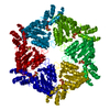
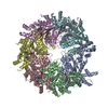
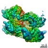
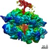
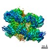
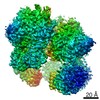
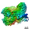
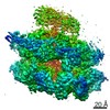
| Title | Structure of ADP-bound N-ethylmaleimide sensitive factor determined by single particle cryoelectron microscopy | |||||||||
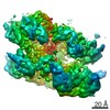
 Map data Map data | Map of ADP-bound N-ethylmaleimide sensitive factor. This map is unsharpened and unfiltered. The map was normalized using the program MAPMAN. | |||||||||
 Sample Sample |
| |||||||||
 Keywords Keywords | ATPases associated with diverse cellular activities | |||||||||
| Function / homology |  Function and homology information Function and homology informationSNARE complex disassembly / ATP-dependent protein disaggregase activity /  vesicle-fusing ATPase / syntaxin-1 binding / positive regulation of receptor recycling / ionotropic glutamate receptor binding / SNARE binding / PDZ domain binding / intracellular protein transport / potassium ion transport ...SNARE complex disassembly / ATP-dependent protein disaggregase activity / vesicle-fusing ATPase / syntaxin-1 binding / positive regulation of receptor recycling / ionotropic glutamate receptor binding / SNARE binding / PDZ domain binding / intracellular protein transport / potassium ion transport / positive regulation of protein catabolic process / midbody / protein-containing complex binding / protein kinase binding / Golgi apparatus / ATP hydrolysis activity / ATP binding / identical protein binding / metal ion binding / plasma membrane / cytosol vesicle-fusing ATPase / syntaxin-1 binding / positive regulation of receptor recycling / ionotropic glutamate receptor binding / SNARE binding / PDZ domain binding / intracellular protein transport / potassium ion transport ...SNARE complex disassembly / ATP-dependent protein disaggregase activity / vesicle-fusing ATPase / syntaxin-1 binding / positive regulation of receptor recycling / ionotropic glutamate receptor binding / SNARE binding / PDZ domain binding / intracellular protein transport / potassium ion transport / positive regulation of protein catabolic process / midbody / protein-containing complex binding / protein kinase binding / Golgi apparatus / ATP hydrolysis activity / ATP binding / identical protein binding / metal ion binding / plasma membrane / cytosolSimilarity search - Function | |||||||||
| Biological species |   Cricetulus griseus (Chinese hamster) Cricetulus griseus (Chinese hamster) | |||||||||
| Method | single particle reconstruction / cryo EM / Resolution: 7.6 Å | |||||||||
 Authors Authors | Zhao M / Wu S / Zhou Q / Vivona S / Cipriano DJ / Cheng Y / Brunger AT | |||||||||
 Citation Citation | Journal: Nature / Year: 2015 Title: Mechanistic insights into the recycling machine of the SNARE complex. Authors: Minglei Zhao / Shenping Wu / Qiangjun Zhou / Sandro Vivona / Daniel J Cipriano / Yifan Cheng / Axel T Brunger /  Abstract: Evolutionarily conserved SNARE (soluble N-ethylmaleimide sensitive factor attachment protein receptors) proteins form a complex that drives membrane fusion in eukaryotes. The ATPase NSF (N- ...Evolutionarily conserved SNARE (soluble N-ethylmaleimide sensitive factor attachment protein receptors) proteins form a complex that drives membrane fusion in eukaryotes. The ATPase NSF (N-ethylmaleimide sensitive factor), together with SNAPs (soluble NSF attachment protein), disassembles the SNARE complex into its protein components, making individual SNAREs available for subsequent rounds of fusion. Here we report structures of ATP- and ADP-bound NSF, and the NSF/SNAP/SNARE (20S) supercomplex determined by single-particle electron cryomicroscopy at near-atomic to sub-nanometre resolution without imposing symmetry. Large, potentially force-generating, conformational differences exist between ATP- and ADP-bound NSF. The 20S supercomplex exhibits broken symmetry, transitioning from six-fold symmetry of the NSF ATPase domains to pseudo four-fold symmetry of the SNARE complex. SNAPs interact with the SNARE complex with an opposite structural twist, suggesting an unwinding mechanism. The interfaces between NSF, SNAPs, and SNAREs exhibit characteristic electrostatic patterns, suggesting how one NSF/SNAP species can act on many different SNARE complexes. | |||||||||
| History |
|
- Structure visualization
| Movie |
Movie viewer |
|---|---|
| Structure viewer | EM map: SurfViewMolmilJmol/JSmol |
| Supplemental images |

- Downloads & links
-EMDB archive
| Map data | emd_6205.map.gz | 6 MB | EMDB map data format | |
|---|---|---|---|---|
| Header (meta data) | emd-6205-v30.xmlemd-6205.xml | 10.7 KB 10.7 KB | Display Display | EMDB header |
| Images | 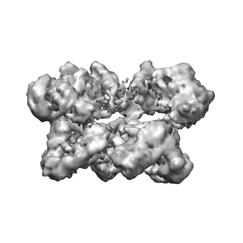 emd_6205.png emd_6205.png | 81.5 KB | ||
| Others | EMD-6205_ADP_NSF_sharpened_-479.map.gz | 7.5 MB | ||
| Archive directory |  http://ftp.pdbj.org/pub/emdb/structures/EMD-6205ftp://ftp.pdbj.org/pub/emdb/structures/EMD-6205 http://ftp.pdbj.org/pub/emdb/structures/EMD-6205ftp://ftp.pdbj.org/pub/emdb/structures/EMD-6205 | HTTPS FTP |
-Related structure data
| Related structure data | 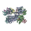 3j95MC 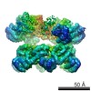 6204C 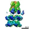 6206C 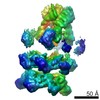 6207C 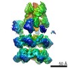 6208C 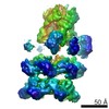 6209C 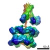 6210C 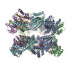 3j94C 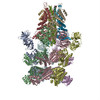 3j96C 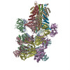 3j97C 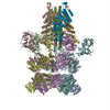 3j98C 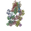 3j99C M: atomic model generated by this map C: citing same article ( |
|---|---|
| Similar structure data |
-Links
| EMDB pages | EMDB (EBI/PDBe) / EMDataResource |
|---|---|
| Related items in Molecule of the Month |


-Map
| File | Download / File: emd_6205.map.gz / Format: CCP4 / Size: 7.8 MB / Type: IMAGE STORED AS FLOATING POINT NUMBER (4 BYTES) | ||||||||||||||||||||||||||||||||||||||||||||||||||||||||||||||||||||
|---|---|---|---|---|---|---|---|---|---|---|---|---|---|---|---|---|---|---|---|---|---|---|---|---|---|---|---|---|---|---|---|---|---|---|---|---|---|---|---|---|---|---|---|---|---|---|---|---|---|---|---|---|---|---|---|---|---|---|---|---|---|---|---|---|---|---|---|---|---|
| Annotation | Map of ADP-bound N-ethylmaleimide sensitive factor. This map is unsharpened and unfiltered. The map was normalized using the program MAPMAN. | ||||||||||||||||||||||||||||||||||||||||||||||||||||||||||||||||||||
| Voxel size | X=Y=Z: 2.4312 Å | ||||||||||||||||||||||||||||||||||||||||||||||||||||||||||||||||||||
| Density |
| ||||||||||||||||||||||||||||||||||||||||||||||||||||||||||||||||||||
| Symmetry | Space group: 1 | ||||||||||||||||||||||||||||||||||||||||||||||||||||||||||||||||||||
| Details | EMDB XML:
CCP4 map header:
| ||||||||||||||||||||||||||||||||||||||||||||||||||||||||||||||||||||
-Supplemental data
-Supplemental map: EMD-6205 ADP NSF sharpened -479.map
| File | EMD-6205_ADP_NSF_sharpened_-479.map | ||||||||||||
|---|---|---|---|---|---|---|---|---|---|---|---|---|---|
| Projections & Slices |
| ||||||||||||
| Density Histograms |
 Z
Z Y
Y X
X


- Sample components
-Entire : ADP-bound N-ethylmaleimide sensitive factor
| Entire | Name: ADP-bound N-ethylmaleimide sensitive factor |
|---|---|
| Components |
|
-Supramolecule #1000: ADP-bound N-ethylmaleimide sensitive factor
| Supramolecule | Name: ADP-bound N-ethylmaleimide sensitive factor / type: sample / ID: 1000 / Oligomeric state: hexamer / Number unique components: 1 |
|---|---|
| Molecular weight | Theoretical: 500 KDa |
-Macromolecule #1: N-ethylmaleimide sensitive factor
| Macromolecule | Name: N-ethylmaleimide sensitive factor / type: protein_or_peptide / ID: 1 / Name.synonym: NSF / Number of copies: 6 / Oligomeric state: hexamer / Recombinant expression: Yes |
|---|---|
| Source (natural) | Organism: Cricetulus griseus (Chinese hamster) / synonym: Chinese hamster |
| Molecular weight | Theoretical: 83 KDa |
| Recombinant expression | Organism:  Escherichia coli (E. coli) / Recombinant strain: BL21(DE3)-RIL / Recombinant plasmid: pPROEX-1 Escherichia coli (E. coli) / Recombinant strain: BL21(DE3)-RIL / Recombinant plasmid: pPROEX-1 |
| Sequence | UniProtKB: Vesicle-fusing ATPase |
-Experimental details
-Structure determination
| Method | cryo EM |
|---|---|
 Processing Processing | single particle reconstruction |
| Aggregation state | particle |
-Sample preparation
| Concentration | 15 mg/mL |
|---|---|
| Buffer | pH: 8 Details: 50 mM Tris-Cl, 150 mM NaCl, 1 mM EDTA, 1 mM ATP, 1 mM DTT, 0.05% v/v Nonident P-40 |
| Grid | Details: Holey carbon on top of 400 mesh copper grid |
| Vitrification | Cryogen name: ETHANE / Chamber humidity: 100 % / Chamber temperature: 90 K / Instrument: FEI VITROBOT MARK I / Method: Blot for 3.5 seconds before plunging. |
- Electron microscopy
| Microscope | FEI POLARA 300 |
|---|---|
| Electron beam | Acceleration voltage: 300 kV / Electron source: FIELD EMISSION GUN |
| Electron optics | Illumination mode: FLOOD BEAM / Imaging mode: BRIGHT FIELDBright-field microscopy / Cs: 2.3 mm / Nominal defocus max: -2.8 µm / Nominal defocus min: -1.8 µm / Nominal magnification: 31000 |
| Sample stage | Specimen holder model: OTHER |
| Date | Jan 14, 2014 |
| Image recording | Category: CCD / Film or detector model: GATAN K2 (4k x 4k) / Average electron dose: 44 e/Å2 Details: Gatan K2 Summit in super-resolution counting mode. Motion correction as described in Li et al. (2013) Nature Methods. |
| Experimental equipment |  Model: Tecnai Polara / Image courtesy: FEI Company |
-Image processing
| CTF correction | Details: Each particle |
|---|---|
| Final reconstruction | Resolution.type: BY AUTHOR / Resolution: 7.6 Å / Resolution method: OTHER / Software - Name: RELION / Number images used: 12830 |
| Details | 3D classification, refinement, and reconstruction were performed using RELION. |
-Atomic model buiding 1
| Initial model | PDB ID: Chain - Chain ID: A |
|---|---|
| Software | Name: Chimera, PHENIX |
| Details | D2 domain of NSF was from crystal structure 1NSF. D1 domain of NSF was from related entry EMD-6204. |
| Refinement | Space: RECIPROCAL / Protocol: FLEXIBLE FIT / Target criteria: R-factor |
| Output model | PDB-3j95: |