 Movie
Movie Controller

[English] 日本語
 Yorodumi
- EMDB-5950: Cryo-electron tomography reveals ciliary defects underlying human... -
+ Open data

- Basic information
| Entry | Database: EMDB / ID: EMD-5950 | |||||||||
|---|---|---|---|---|---|---|---|---|---|---|
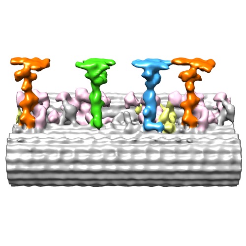
| Title | Cryo-electron tomography reveals ciliary defects underlying human RSPH1 primary ciliary dyskinesia | |||||||||
 Map data Map data | Reconstruction of 96 nm axonemal repeat of normal human respiratory cilia | |||||||||
 Sample Sample |
| |||||||||
 Keywords Keywords |  axoneme / ciliopathy / human respiratory cilia / radial spoke / dynein axoneme / ciliopathy / human respiratory cilia / radial spoke / dynein | |||||||||
| Biological species |  Homo sapiens (human) Homo sapiens (human) | |||||||||
| Method | subtomogram averaging / cryo EM / Resolution: 34.0 Å | |||||||||
 Authors Authors | Lin J / Yin W / Smith MC / Song KK / Leigh MW / Zariwala MA / Knowles MR / Ostrowski LE / Nicastro D | |||||||||
 Citation Citation | Journal: Nat Commun / Year: 2014 Title: Cryo-electron tomography reveals ciliary defects underlying human RSPH1 primary ciliary dyskinesia. Authors: Jianfeng Lin / Weining Yin / Maria C Smith / Kangkang Song / Margaret W Leigh / Maimoona A Zariwala / Michael R Knowles / Lawrence E Ostrowski / Daniela Nicastro /  Abstract: Cilia play essential roles in normal human development and health; cilia dysfunction results in diseases such as primary ciliary dyskinesia (PCD). Despite their importance, the native structure of ...Cilia play essential roles in normal human development and health; cilia dysfunction results in diseases such as primary ciliary dyskinesia (PCD). Despite their importance, the native structure of human cilia is unknown, and structural defects in the cilia of patients are often undetectable or remain elusive because of heterogeneity. Here we develop an approach that enables visualization of human (patient) cilia at high-resolution using cryo-electron tomography of samples obtained noninvasively by nasal scrape biopsy. We present the native 3D structures of normal and PCD-causing RSPH1-mutant human respiratory cilia in unprecedented detail; this allows comparisons of cilia structure across evolutionarily distant species and reveals the previously unknown primary defect and the heterogeneous secondary defects in RSPH1-mutant cilia. Our data provide evidence for structural and functional heterogeneity in radial spokes, suggest a mechanism for the milder RSPH1 PCD phenotype and demonstrate that cryo-electron tomography can be applied to human disease by directly imaging patient samples. | |||||||||
| History |
|
- Structure visualization
| Movie |
 Movie viewer Movie viewer |
|---|---|
| Structure viewer | EM map: SurfViewMolmilJmol/JSmol |
| Supplemental images |
- Downloads & links
-EMDB archive
| Map data | emd_5950.map.gz | 3.1 MB | EMDB map data format | |
|---|---|---|---|---|
| Header (meta data) | emd-5950-v30.xmlemd-5950.xml | 9.9 KB 9.9 KB | Display Display | EMDB header |
| Images | emd_5950.tif | 173.3 KB | ||
| Archive directory |  http://ftp.pdbj.org/pub/emdb/structures/EMD-5950ftp://ftp.pdbj.org/pub/emdb/structures/EMD-5950 http://ftp.pdbj.org/pub/emdb/structures/EMD-5950ftp://ftp.pdbj.org/pub/emdb/structures/EMD-5950 | HTTPS FTP |
-Related structure data
| Similar structure data |
|---|
-Links
| EMDB pages | EMDB (EBI/PDBe) / EMDataResource |
|---|
-Map
| File | Download / File: emd_5950.map.gz / Format: CCP4 / Size: 3.9 MB / Type: IMAGE STORED AS FLOATING POINT NUMBER (4 BYTES) | ||||||||||||||||||||||||||||||||||||||||||||||||||||||||||||||||||||
|---|---|---|---|---|---|---|---|---|---|---|---|---|---|---|---|---|---|---|---|---|---|---|---|---|---|---|---|---|---|---|---|---|---|---|---|---|---|---|---|---|---|---|---|---|---|---|---|---|---|---|---|---|---|---|---|---|---|---|---|---|---|---|---|---|---|---|---|---|---|
| Annotation | Reconstruction of 96 nm axonemal repeat of normal human respiratory cilia | ||||||||||||||||||||||||||||||||||||||||||||||||||||||||||||||||||||
| Voxel size | X=Y=Z: 9.997 Å | ||||||||||||||||||||||||||||||||||||||||||||||||||||||||||||||||||||
| Density |
| ||||||||||||||||||||||||||||||||||||||||||||||||||||||||||||||||||||
| Symmetry | Space group: 1 | ||||||||||||||||||||||||||||||||||||||||||||||||||||||||||||||||||||
| Details | EMDB XML:
CCP4 map header:
| ||||||||||||||||||||||||||||||||||||||||||||||||||||||||||||||||||||
-Supplemental data
- Sample components
-Entire : Normal human respiratory ciliary axonemes
| Entire | Name: Normal human respiratory ciliary axonemes |
|---|---|
| Components |
|
-Supramolecule #1000: Normal human respiratory ciliary axonemes
| Supramolecule | Name: Normal human respiratory ciliary axonemes / type: sample / ID: 1000 Details: The samples were normal human respiratory ciliary axonemes isolated from cultures of trachea-bronchial epithelial cells. Number unique components: 1 |
|---|
-Supramolecule #1: Axoneme
| Supramolecule | Name: Axoneme / type: organelle_or_cellular_component / ID: 1 / Recombinant expression: No / Database: NCBI |
|---|---|
| Source (natural) | Organism: Homo sapiens (human) / synonym: Human / Tissue: Trachea-bronchial / Cell: Epithelial cells / Organelle: Cilia |
-Experimental details
-Structure determination
| Method | cryo EM |
|---|---|
 Processing Processing | subtomogram averaging |
| Aggregation state | cell |
-Sample preparation
| Buffer | pH: 7.3 Details: 30 mM Hepes, pH 7.3, 1 mM EGTA, 5 mM MgSO4, 0.1 mM EDTA, 25 mM NaCl, 1 mM dithiothreitol, 1% protease inhibitor cocktail, 100 g/mL soybean trypsin inhibitor (Sigma T9128) |
|---|---|
| Grid | Details: Quantifoil holey carbon grids Cu 200 mesh R2/2 |
| Vitrification | Cryogen name: ETHANE / Chamber temperature: 100 K / Instrument: HOMEMADE PLUNGER / Method: Blot for 1.5-2.5 seconds before plunging |
- Electron microscopy
| Microscope | FEI TECNAI F30 |
|---|---|
| Electron beam | Acceleration voltage: 300 kV / Electron source: FIELD EMISSION GUN |
| Electron optics | Illumination mode: FLOOD BEAM / Imaging mode: BRIGHT FIELDBright-field microscopy / Nominal defocus max: 8.0 µm / Nominal defocus min: 6.0 µm / Nominal magnification: 13500 |
| Specialist optics | Energy filter - Name: GATAN postcolumn filter GIF / Energy filter - Lower energy threshold: 0.0 eV / Energy filter - Upper energy threshold: 20.0 eV |
| Sample stage | Specimen holder model: GATAN LIQUID NITROGEN / Tilt series - Axis1 - Min angle: -65 ° / Tilt series - Axis1 - Max angle: 65 ° |
| Date | Feb 5, 2013 |
| Image recording | Category: CCD / Film or detector model: GATAN ULTRASCAN 1000 (2k x 2k) / Average electron dose: 100 e/Å2 |
| Experimental equipment |  Model: Tecnai F30 / Image courtesy: FEI Company |
-Image processing
| Final reconstruction | Algorithm: OTHER / Resolution.type: BY AUTHOR / Resolution: 34.0 Å / Resolution method: OTHER / Software - Name: IMOD Details: Final map was calculated by averaging 850 particles (96-nm-long axonemal repeats) from 12 tomograms. Number subtomograms used: 950 |
|---|---|
| Details | 3D tomograms were reconstructed using fiducial alignment and weighted backprojection using IMOD software. Final maps were calculated by averaging 950 particles (96-nm-long axonemal repeats) from 12 tomograms using the PEET software. |
