 Movie
Movie Controller

[English] 日本語
 Yorodumi
- EMDB-5922: 3.6 Angstrom resolution MAVS filament generated from helical reco... -
+ Open data

- Basic information
| Entry | Database: EMDB / ID: EMD-5922 | |||||||||
|---|---|---|---|---|---|---|---|---|---|---|
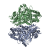
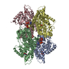
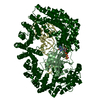
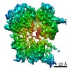
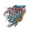
| Title | 3.6 Angstrom resolution MAVS filament generated from helical reconstruction | |||||||||
 Map data Map data | 3.6 Angstrom resolution MAVS filament generated from helical reconstruction | |||||||||
 Sample Sample |
| |||||||||
 Keywords Keywords | CARD / MAVS /  innate immunity / RIG-I / MDA5 / spontaneous filament formation innate immunity / RIG-I / MDA5 / spontaneous filament formation | |||||||||
| Function / homology |  Function and homology information Function and homology informationpositive regulation of IP-10 production / regulation of peroxisome organization / RIG-I binding / positive regulation of chemokine (C-C motif) ligand 5 production / positive regulation of myeloid dendritic cell cytokine production / CARD domain binding / NF-kB activation through FADD/RIP-1 pathway mediated by caspase-8 and -10 / positive regulation of response to cytokine stimulus / protein localization to mitochondrion / positive regulation of type I interferon-mediated signaling pathway ...positive regulation of IP-10 production / regulation of peroxisome organization / RIG-I binding / positive regulation of chemokine (C-C motif) ligand 5 production / positive regulation of myeloid dendritic cell cytokine production / CARD domain binding / NF-kB activation through FADD/RIP-1 pathway mediated by caspase-8 and -10 / positive regulation of response to cytokine stimulus / protein localization to mitochondrion / positive regulation of type I interferon-mediated signaling pathway / peroxisomal membrane / TRAF6 mediated IRF7 activation / negative regulation of type I interferon-mediated signaling pathway / positive regulation of NLRP3 inflammasome complex assembly / negative regulation of viral genome replication / cellular response to exogenous dsRNA / cytoplasmic pattern recognition receptor signaling pathway / type I interferon-mediated signaling pathway / positive regulation of interferon-alpha production / antiviral innate immune response / TRAF6 mediated NF-kB activation / positive regulation of type I interferon production / ubiquitin ligase complex / signaling adaptor activity / positive regulation of defense response to virus by host / molecular condensate scaffold activity / activation of innate immune response / positive regulation of interferon-beta production / Negative regulators of DDX58/IFIH1 signaling / positive regulation of interleukin-8 production / mitochondrial membrane / DDX58/IFIH1-mediated induction of interferon-alpha/beta / PKR-mediated signaling / positive regulation of interleukin-6 production / positive regulation of protein import into nucleus / SARS-CoV-1 activates/modulates innate immune responses / positive regulation of DNA-binding transcription factor activity / Ovarian tumor domain proteases / positive regulation of tumor necrosis factor production / TRAF3-dependent IRF activation pathway / defense response to virus / positive regulation of canonical NF-kappaB signal transduction / mitochondrial outer membrane / molecular adaptor activity / defense response to bacterium / positive regulation of protein phosphorylation / innate immune response / protein kinase binding / SARS-CoV-2 activates/modulates innate and adaptive immune responses / signal transduction / positive regulation of transcription by RNA polymerase II / mitochondrion / identical protein bindingSimilarity search - Function | |||||||||
| Biological species |  Homo sapiens (human) Homo sapiens (human) | |||||||||
| Method | helical reconstruction / cryo EM / Resolution: 3.64 Å | |||||||||
 Authors Authors | Wu B / Peisley A / Li Z / Egelman E / Walz T / Penczek P / Hur S | |||||||||
 Citation Citation | Journal: Mol Cell / Year: 2014 Title: Molecular imprinting as a signal-activation mechanism of the viral RNA sensor RIG-I. Authors: Bin Wu / Alys Peisley / David Tetrault / Zongli Li / Edward H Egelman / Katharine E Magor / Thomas Walz / Pawel A Penczek / Sun Hur /   Abstract: RIG-I activates interferon signaling pathways by promoting filament formation of the adaptor molecule, MAVS. Assembly of the MAVS filament is mediated by its CARD domain (CARD(MAVS)), and requires ...RIG-I activates interferon signaling pathways by promoting filament formation of the adaptor molecule, MAVS. Assembly of the MAVS filament is mediated by its CARD domain (CARD(MAVS)), and requires its interaction with the tandem CARDs of RIG-I (2CARD(RIG-I)). However, the precise nature of the interaction between 2CARD(RIG-I) and CARD(MAVS), and how this interaction leads to CARD(MAVS) filament assembly, has been unclear. Here we report a 3.6 Å electron microscopy structure of the CARD(MAVS) filament and a 3.4 Å crystal structure of the 2CARD(RIG-I):CARD(MAVS) complex, representing 2CARD(RIG-I) "caught in the act" of nucleating the CARD(MAVS) filament. These structures, together with functional analyses, show that 2CARD(RIG-I) acts as a template for the CARD(MAVS) filament assembly, by forming a helical tetrameric structure and recruiting CARD(MAVS) along its helical trajectory. Our work thus reveals that signal activation by RIG-I occurs by imprinting its helical assembly architecture on MAVS, a previously uncharacterized mechanism of signal transmission. | |||||||||
| History |
|
- Structure visualization
| Movie |
Movie viewer |
|---|---|
| Structure viewer | EM map: SurfViewMolmilJmol/JSmol |
| Supplemental images |

- Downloads & links
-EMDB archive
| Map data | emd_5922.map.gz | 8.1 MB | EMDB map data format | |
|---|---|---|---|---|
| Header (meta data) | emd-5922-v30.xmlemd-5922.xml | 9.5 KB 9.5 KB | Display Display | EMDB header |
| Images |  400_5922.gif 400_5922.gif 80_5922.gif 80_5922.gif | 95.5 KB 5.9 KB | ||
| Archive directory |  http://ftp.pdbj.org/pub/emdb/structures/EMD-5922ftp://ftp.pdbj.org/pub/emdb/structures/EMD-5922 http://ftp.pdbj.org/pub/emdb/structures/EMD-5922ftp://ftp.pdbj.org/pub/emdb/structures/EMD-5922 | HTTPS FTP |
-Related structure data


-Links
| EMDB pages | EMDB (EBI/PDBe) / EMDataResource |
|---|---|
| Related items in Molecule of the Month |


-Map
| File | Download / File: emd_5922.map.gz / Format: CCP4 / Size: 12.2 MB / Type: IMAGE STORED AS FLOATING POINT NUMBER (4 BYTES) | ||||||||||||||||||||||||||||||||||||||||||||||||||||||||||||||||||||
|---|---|---|---|---|---|---|---|---|---|---|---|---|---|---|---|---|---|---|---|---|---|---|---|---|---|---|---|---|---|---|---|---|---|---|---|---|---|---|---|---|---|---|---|---|---|---|---|---|---|---|---|---|---|---|---|---|---|---|---|---|---|---|---|---|---|---|---|---|---|
| Annotation | 3.6 Angstrom resolution MAVS filament generated from helical reconstruction | ||||||||||||||||||||||||||||||||||||||||||||||||||||||||||||||||||||
| Voxel size | X=Y=Z: 0.62 Å | ||||||||||||||||||||||||||||||||||||||||||||||||||||||||||||||||||||
| Density |
| ||||||||||||||||||||||||||||||||||||||||||||||||||||||||||||||||||||
| Symmetry | Space group: 1 | ||||||||||||||||||||||||||||||||||||||||||||||||||||||||||||||||||||
| Details | EMDB XML:
CCP4 map header:
| ||||||||||||||||||||||||||||||||||||||||||||||||||||||||||||||||||||
-Supplemental data
- Sample components
-Entire : MAVS filament
| Entire | Name: MAVS filament |
|---|---|
| Components |
|
-Supramolecule #1000: MAVS filament
| Supramolecule | Name: MAVS filament / type: sample / ID: 1000 / Details: The density is only a section of the filament. / Oligomeric state: filament / Number unique components: 1 |
|---|---|
| Molecular weight | Experimental: 150 KDa / Theoretical: 150 KDa / Method: Size exclusion chromatography |
-Macromolecule #1: Mitochondria Anti-viral Signaling protein, CARD domain
| Macromolecule | Name: Mitochondria Anti-viral Signaling protein, CARD domain type: protein_or_peptide / ID: 1 / Name.synonym: MAVS / Oligomeric state: filament / Recombinant expression: Yes |
|---|---|
| Source (natural) | Organism: Homo sapiens (human) / synonym: human |
| Recombinant expression | Organism:  Escherichia coli (E. coli) / Recombinant strain: BL21(DE3) Escherichia coli (E. coli) / Recombinant strain: BL21(DE3) |
| Sequence | UniProtKB: Mitochondrial antiviral-signaling protein |
-Experimental details
-Structure determination
| Method | cryo EM |
|---|---|
 Processing Processing | helical reconstruction |
| Aggregation state | filament |
-Sample preparation
| Concentration | 0.2 mg/mL |
|---|---|
| Buffer | pH: 7.5 / Details: 20 mM Tris, 150 mM NaCl |
| Grid | Details: glow-discharged Quantifoil R1.2/1.3 holey carbon grids |
| Vitrification | Cryogen name: NITROGEN / Instrument: FEI VITROBOT MARK I |
- Electron microscopy
| Microscope | FEI TECNAI F20 |
|---|---|
| Electron beam | Acceleration voltage: 200 kV / Electron source: FIELD EMISSION GUN |
| Electron optics | Calibrated magnification: 40410 / Illumination mode: FLOOD BEAM / Imaging mode: BRIGHT FIELDBright-field microscopy / Nominal magnification: 29000 |
| Specialist optics | Energy filter - Name: FEI |
| Sample stage | Specimen holder model: GATAN LIQUID NITROGEN |
| Details | Movies were recorded at liquid nitrogen temperature with a K2 Summit direct detector device camera operated in super-resolution mode with dose-fractionation. |
| Date | Aug 10, 2013 |
| Image recording | Category: CCD / Film or detector model: GATAN K2 (4k x 4k) / Number real images: 1863 / Average electron dose: 31 e/Å2 |
| Experimental equipment |  Model: Tecnai F20 / Image courtesy: FEI Company |
-Image processing
| Final reconstruction | Applied symmetry - Helical parameters - Δz: 5.13 Å Applied symmetry - Helical parameters - Δ&Phi: 101.1 ° Applied symmetry - Helical parameters - Axial symmetry: C1 (asymmetric) Resolution.type: BY AUTHOR / Resolution: 3.64 Å / Resolution method: OTHER / Software - Name: Helicon |
|---|---|
| Details | The electron density map of the filament was reconstructed using a helical geometrically constrained reconstruction approach (Helicon). |
