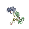
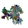
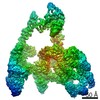
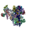
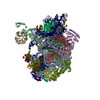
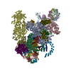
Journal: Cell / Year: 2013 Title: Structure of the mammalian ribosomal 43S preinitiation complex bound to the scanning factor DHX29. Authors: Yaser Hashem / Amedee des Georges / Vidya Dhote / Robert Langlois / Hstau Y Liao / Robert A Grassucci / Christopher U T Hellen / Tatyana V Pestova / Joachim Frank / Abstract: Eukaryotic translation initiation begins with assembly of a 43S preinitiation complex. First, methionylated initiator methionine transfer RNA (Met-tRNAi(Met)), eukaryotic initiation factor (eIF) 2, ...Eukaryotic translation initiation begins with assembly of a 43S preinitiation complex. First, methionylated initiator methionine transfer RNA (Met-tRNAi(Met)), eukaryotic initiation factor (eIF) 2, and guanosine triphosphate form a ternary complex (TC). The TC, eIF3, eIF1, and eIF1A cooperatively bind to the 40S subunit, yielding the 43S preinitiation complex, which is ready to attach to messenger RNA (mRNA) and start scanning to the initiation codon. Scanning on structured mRNAs additionally requires DHX29, a DExH-box protein that also binds directly to the 40S subunit. Here, we present a cryo-electron microscopy structure of the mammalian DHX29-bound 43S complex at 11.6 Å resolution. It reveals that eIF2 interacts with the 40S subunit via its α subunit and supports Met-tRNAi(Met) in an unexpected P/I orientation (eP/I). The structural core of eIF3 resides on the back of the 40S subunit, establishing two principal points of contact, whereas DHX29 binds around helix 16. The structure provides insights into eukaryote-specific aspects of translation, including the mechanism of action of DHX29.
History
Deposition
May 1, 2013
-
Header (metadata) release
May 22, 2013
-
Map release
Jun 5, 2013
-
Update
Jun 5, 2013
-
Current status
Jun 5, 2013
Processing site: RCSB / Status: Released
-
Structure visualization
Movie
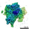
Surface view with section colored by density value
Download / File: emd_5658.map.gz / Format: CCP4 / Size: 47.7 MB / Type: IMAGE STORED AS FLOATING POINT NUMBER (4 BYTES)
Annotation
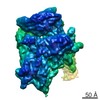
Reconstruction of the mammalian 43S preinitiation complex bound to DHX29
Voxel size
X=Y=Z: 2.245 Å
Density
Contour Level
By AUTHOR: 0.019 / Movie #1: 0.019
Minimum - Maximum
-0.02738628 - 0.11589623
Average (Standard dev.)
0.00035334 (±0.00863221)
Symmetry
Space group: 1
Details
EMDB XML:
Map geometry
Axis order
X
Y
Z
Origin
0
0
0
Dimensions
234
234
234
Spacing
234
234
234
Cell
A=B=C: 525.32996 Å α=β=γ: 90.0 °
CCP4 map header:
mode
Image stored as Reals
Å/pix. X/Y/Z
2.245
2.245
2.245
M x/y/z
234
234
234
origin x/y/z
0.000
0.000
0.000
length x/y/z
525.330
525.330
525.330
α/β/γ
90.000
90.000
90.000
start NX/NY/NZ
-132
-122
-147
NX/NY/NZ
250
274
261
MAP C/R/S
1
2
3
start NC/NR/NS
0
0
0
NC/NR/NS
234
234
234
D min/max/mean
-0.027
0.116
0.000
-
Supplemental data
-
Sample components
-
Entire : Cryo-electron microscopy structure of the mammalian 43S preinitia...
Entire
Name: Cryo-electron microscopy structure of the mammalian 43S preinitiation complex bound to DHX29
Components
Sample: Cryo-electron microscopy structure of the mammalian 43S preinitiation complex bound to DHX29
Complex: eukaryotic small ribosomal subunit
Protein or peptide: eukaryotic initiation factor 3
Protein or peptide: eukaryotic initiation factor 2
Protein or peptide: eukaryotic initiation factor 1
Protein or peptide: eukaryotic initiation factor 1A
Protein or peptide: DHX29
RNA: Transfer RNA
-
Supramolecule #1000: Cryo-electron microscopy structure of the mammalian 43S preinitia...
Supramolecule
Name: Cryo-electron microscopy structure of the mammalian 43S preinitiation complex bound to DHX29 type: sample / ID: 1000 Oligomeric state: One 40S binds one eIF3, one eIF2, one Met-tRNAiMet, and one DHX29 Number unique components: 5
-
Supramolecule #1: eukaryotic small ribosomal subunit
Cryogen name: ETHANE / Chamber humidity: 100 % / Chamber temperature: 120 K / Instrument: FEI VITROBOT MARK II / Method: Blot for seconds before plunging
-
Electron microscopy
Microscope
FEI TECNAI 20
Temperature
Average: 110 K
Date
Nov 1, 2012
Image recording
Category: CCD / Film or detector model: GATAN ULTRASCAN 4000 (4k x 4k) / Number real images: 8000 / Average electron dose: 12 e/Å2 / Bits/pixel: 32
Electron beam
Acceleration voltage: 110 kV / Electron source: FIELD EMISSION GUN
The particles of this reconstruction were obtained after particle sorting using Relion
CTF correction
Details: Each particle
Final reconstruction
Algorithm: OTHER / Resolution.type: BY AUTHOR / Resolution: 11.6 Å / Resolution method: OTHER / Software - Name: Spider, Relion / Number images used: 29000
In the structure databanks used in Yorodumi, some data are registered as the other names, "COVID-19 virus" and "2019-nCoV". Here are the details of the virus and the list of structure data.
Jan 31, 2019. EMDB accession codes are about to change! (news from PDBe EMDB page)
EMDB accession codes are about to change! (news from PDBe EMDB page)
The allocation of 4 digits for EMDB accession codes will soon come to an end. Whilst these codes will remain in use, new EMDB accession codes will include an additional digit and will expand incrementally as the available range of codes is exhausted. The current 4-digit format prefixed with “EMD-” (i.e. EMD-XXXX) will advance to a 5-digit format (i.e. EMD-XXXXX), and so on. It is currently estimated that the 4-digit codes will be depleted around Spring 2019, at which point the 5-digit format will come into force.
The EM Navigator/Yorodumi systems omit the EMD- prefix.
Related info.:Q: What is EMD? / ID/Accession-code notation in Yorodumi/EM Navigator
Yorodumi is a browser for structure data from EMDB, PDB, SASBDB, etc.
This page is also the successor to EM Navigator detail page, and also detail information page/front-end page for Omokage search.
The word "yorodu" (or yorozu) is an old Japanese word meaning "ten thousand". "mi" (miru) is to see.
Related info.:EMDB / PDB / SASBDB / Comparison of 3 databanks / Yorodumi Search / Aug 31, 2016. New EM Navigator & Yorodumi / Yorodumi Papers / Jmol/JSmol / Function and homology information / Changes in new EM Navigator and Yorodumi
 Movie
Movie Controller

 Yorodumi
 Open data
 Basic information
 Map data
 Sample
 Keywords
 Function and homology information
 Authors
 Citation
 Structure visualization
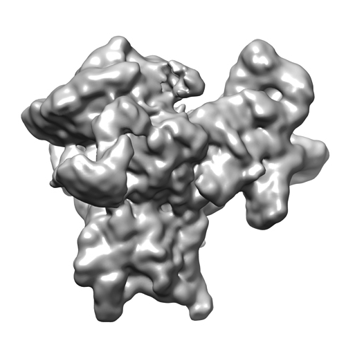
 Downloads & links
 emd_5658_1.jpg
 http://ftp.pdbj.org/pub/emdb/structures/EMD-5658
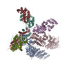


 Sample components
 Processing
 Electron microscopy
 FIELD EMISSION GUN