 Movie
Movie Controller


+ Open data

- Basic information
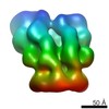
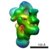
| Entry | Database: EMDB / ID: EMD-5638 | |||||||||
|---|---|---|---|---|---|---|---|---|---|---|
| Title | 3D Cryo-negative EM structure of nucleosome-bound SWR1 | |||||||||
 Map data Map data | Cryo-EM structure of SWR1 (S.c.) bound to recombinant nucleosomes | |||||||||
 Sample Sample |
| |||||||||
 Keywords Keywords |  chromatin remodeling / SWR1 / INO80 / nucleosome / Rvb1 / Rvb2 / AAA+ ATPase / histone / dimer exchange / H2A.Z chromatin remodeling / SWR1 / INO80 / nucleosome / Rvb1 / Rvb2 / AAA+ ATPase / histone / dimer exchange / H2A.Z | |||||||||
| Biological species |  Saccharomyces cerevisiae (brewer's yeast) Saccharomyces cerevisiae (brewer's yeast) | |||||||||
| Method | single particle reconstruction / cryo EM / negative staining / Resolution: 34.0 Å | |||||||||
 Authors Authors | Nguyen VQ / Ranjan A / Stengel F / Wei D / Aebersold R / Wu C / Leschziner AE | |||||||||
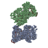
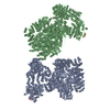
 Citation Citation | Journal: Cell / Year: 2013 Title: Molecular architecture of the ATP-dependent chromatin-remodeling complex SWR1. Authors: Vu Q Nguyen / Anand Ranjan / Florian Stengel / Debbie Wei / Ruedi Aebersold / Carl Wu / Andres E Leschziner /  Abstract: The ATP-dependent chromatin-remodeling complex SWR1 exchanges a variant histone H2A.Z/H2B dimer for a canonical H2A/H2B dimer at nucleosomes flanking histone-depleted regions, such as promoters. This ...The ATP-dependent chromatin-remodeling complex SWR1 exchanges a variant histone H2A.Z/H2B dimer for a canonical H2A/H2B dimer at nucleosomes flanking histone-depleted regions, such as promoters. This localization of H2A.Z is conserved throughout eukaryotes. SWR1 is a 1 megadalton complex containing 14 different polypeptides, including the AAA+ ATPases Rvb1 and Rvb2. Using electron microscopy, we obtained the three-dimensional structure of SWR1 and mapped its major functional components. Our data show that SWR1 contains a single heterohexameric Rvb1/Rvb2 ring that, together with the catalytic subunit Swr1, brackets two independently assembled multisubunit modules. We also show that SWR1 undergoes a large conformational change upon engaging a limited region of the nucleosome core particle. Our work suggests an important structural role for the Rvbs and a distinct substrate-handling mode by SWR1, thereby providing a structural framework for understanding the complex dimer-exchange reaction. | |||||||||
| History |
|
- Structure visualization
| Movie |
 Movie viewer Movie viewer |
|---|---|
| Structure viewer | EM map: SurfViewMolmilJmol/JSmol |
| Supplemental images |

- Downloads & links
-EMDB archive
| Map data | emd_5638.map.gz | 11.6 MB | EMDB map data format | |
|---|---|---|---|---|
| Header (meta data) | emd-5638-v30.xmlemd-5638.xml | 10 KB 10 KB | Display Display | EMDB header |
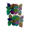
| Images |  emd_5638.png emd_5638.png | 65.6 KB | ||
| Archive directory |  http://ftp.pdbj.org/pub/emdb/structures/EMD-5638ftp://ftp.pdbj.org/pub/emdb/structures/EMD-5638 http://ftp.pdbj.org/pub/emdb/structures/EMD-5638ftp://ftp.pdbj.org/pub/emdb/structures/EMD-5638 | HTTPS FTP |
-Related structure data


-Links
| EMDB pages | EMDB (EBI/PDBe) / EMDataResource |
|---|
-Map
| File | Download / File: emd_5638.map.gz / Format: CCP4 / Size: 12.6 MB / Type: IMAGE STORED AS FLOATING POINT NUMBER (4 BYTES) | ||||||||||||||||||||||||||||||||||||||||||||||||||||||||||||||||||||
|---|---|---|---|---|---|---|---|---|---|---|---|---|---|---|---|---|---|---|---|---|---|---|---|---|---|---|---|---|---|---|---|---|---|---|---|---|---|---|---|---|---|---|---|---|---|---|---|---|---|---|---|---|---|---|---|---|---|---|---|---|---|---|---|---|---|---|---|---|---|
| Annotation | Cryo-EM structure of SWR1 (S.c.) bound to recombinant nucleosomes | ||||||||||||||||||||||||||||||||||||||||||||||||||||||||||||||||||||
| Voxel size | X=Y=Z: 3.45 Å | ||||||||||||||||||||||||||||||||||||||||||||||||||||||||||||||||||||
| Density |
| ||||||||||||||||||||||||||||||||||||||||||||||||||||||||||||||||||||
| Symmetry | Space group: 1 | ||||||||||||||||||||||||||||||||||||||||||||||||||||||||||||||||||||
| Details | EMDB XML:
CCP4 map header:
| ||||||||||||||||||||||||||||||||||||||||||||||||||||||||||||||||||||
-Supplemental data
- Sample components
-Entire : SWR1 (S.c.) bound to recombinant nucleosomes
| Entire | Name: SWR1 (S.c.) bound to recombinant nucleosomes |
|---|---|
| Components |
|
-Supramolecule #1000: SWR1 (S.c.) bound to recombinant nucleosomes
| Supramolecule | Name: SWR1 (S.c.) bound to recombinant nucleosomes / type: sample / ID: 1000 / Number unique components: 15 |
|---|---|
| Molecular weight | Theoretical: 1.2 MDa |
-Macromolecule #1: SWR1
| Macromolecule | Name: SWR1 / type: protein_or_peptide / ID: 1 / Recombinant expression: No / Database: NCBI |
|---|---|
| Source (natural) | Organism: Saccharomyces cerevisiae (brewer's yeast) / Strain: W1588C-4C / synonym: Baker's yeast |
| Molecular weight | Theoretical: 1.2 MDa |
-Experimental details
-Structure determination
| Method | negative staining, cryo EM |
|---|---|
 Processing Processing | single particle reconstruction |
| Aggregation state | particle |
-Sample preparation
| Buffer | pH: 7.4 Details: 25 mM HEPES-KOH, pH 7.6, 1 mM EDTA, 2 mM MgCl2, 0.01% NP-40, 1 mM DTT, 100 mM KCl |
|---|---|
| Staining | Type: NEGATIVE Details: Cryo-negative staining with 2% uranyl formate followed by freezing in liquid nitrogen |
| Grid | Details: 200 mesh Quantifoil with glow-discharged thin carbon support |
| Vitrification | Cryogen name: NITROGEN / Instrument: OTHER Method: Cryo-negative stain with 2% uranyl formate. Blotted and frozen in liquid nitrogen. |
- Electron microscopy
| Microscope | FEI TECNAI F20 |
|---|---|
| Electron beam | Acceleration voltage: 120 kV / Electron source: FIELD EMISSION GUN |
| Electron optics | Calibrated magnification: 86700 / Illumination mode: FLOOD BEAM / Imaging mode: BRIGHT FIELDBright-field microscopy / Cs: 2.0 mm / Nominal defocus max: 2.0 µm / Nominal defocus min: 0.5 µm / Nominal magnification: 62000 |
| Sample stage | Specimen holder model: GATAN LIQUID NITROGEN |
| Temperature | Average: 100 K |
| Details | Low-dose imaging |
| Date | May 20, 2012 |
| Image recording | Category: CCD / Film or detector model: GATAN ULTRASCAN 4000 (4k x 4k) / Digitization - Sampling interval: 15 µm / Number real images: 300 / Average electron dose: 20 e/Å2 |
| Experimental equipment |  Model: Tecnai F20 / Image courtesy: FEI Company |
-Image processing
| CTF correction | Details: EMAN2 |
|---|---|
| Final reconstruction | Algorithm: OTHER / Resolution.type: BY AUTHOR / Resolution: 34.0 Å / Resolution method: OTHER / Software - Name: RELION / Number images used: 12000 |
| Details | The dataset was classified using the maximum-likelihood based method in the RELION program. A selected 3D class was then refined against particles assigned to the class using RELION's "Autorefine" function. |
