 Movie
Movie Controller

[English] 日本語
 Yorodumi
- EMDB-5523: Protease Cleavage Leads to Formation of Mature Trimer Interface i... -
+ Open data

- Basic information
| Entry | Database: EMDB / ID: EMD-5523 | |||||||||
|---|---|---|---|---|---|---|---|---|---|---|
| Title | Protease Cleavage Leads to Formation of Mature Trimer Interface in HIV-1 Capsid | |||||||||
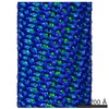
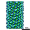
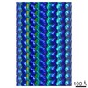
 Map data Map data | Reconstruction of CA in HIV-1 CA-SP1-NC assembly | |||||||||
 Sample Sample |
| |||||||||
 Keywords Keywords | HIV-1 / capsid / nucleocapsid / maturation / cryoEM / cross-linking / interface | |||||||||
| Biological species |   Human immunodeficiency virus 1 Human immunodeficiency virus 1 | |||||||||
| Method | helical reconstruction / cryo EM / Resolution: 13.0 Å | |||||||||
 Authors Authors | Meng X / Zhao G / Yufenyuy E / Ke D / Ning J / DeLucia M / Ahn J / Gronenborn AM / Aiken C / Zhang P | |||||||||
 Citation Citation | Journal: PLoS Pathog / Year: 2012 Title: Protease cleavage leads to formation of mature trimer interface in HIV-1 capsid. Authors: Xin Meng / Gongpu Zhao / Ernest Yufenyuy / Danxia Ke / Jiying Ning / Maria Delucia / Jinwoo Ahn / Angela M Gronenborn / Christopher Aiken / Peijun Zhang /  Abstract: During retrovirus particle maturation, the assembled Gag polyprotein is cleaved by the viral protease into matrix (MA), capsid (CA), and nucleocapsid (NC) proteins. To form the mature viral capsid, ...During retrovirus particle maturation, the assembled Gag polyprotein is cleaved by the viral protease into matrix (MA), capsid (CA), and nucleocapsid (NC) proteins. To form the mature viral capsid, CA rearranges, resulting in a lattice composed of hexameric and pentameric CA units. Recent structural studies of assembled HIV-1 CA revealed several inter-subunit interfaces in the capsid lattice, including a three-fold interhexamer interface that is critical for proper capsid stability. Although a general architecture of immature particles has been provided by cryo-electron tomographic studies, the structural details of the immature particle and the maturation pathway remain unknown. Here, we used cryo-electron microscopy (cryoEM) to determine the structure of tubular assemblies of the HIV-1 CA-SP1-NC protein. Relative to the mature assembled CA structure, we observed a marked conformational difference in the position of the CA-CTD relative to the NTD in the CA-SP1-NC assembly, involving the flexible hinge connecting the two domains. This difference was verified via engineered disulfide crosslinking, revealing that inter-hexamer contacts, in particular those at the pseudo three-fold axis, are altered in the CA-SP1-NC assemblies compared to the CA assemblies. Results from crosslinking analyses of mature and immature HIV-1 particles containing the same Cys substitutions in the Gag protein are consistent with these findings. We further show that cleavage of preassembled CA-SP1-NC by HIV-1 protease in vitro leads to release of SP1 and NC without disassembly of the lattice. Collectively, our results indicate that the proteolytic cleavage of Gag leads to a structural reorganization of the polypeptide and creates the three-fold interhexamer interface, important for the formation of infectious HIV-1 particles. | |||||||||
| History |
|
- Structure visualization
| Movie |
 Movie viewer Movie viewer |
|---|---|
| Structure viewer | EM map: SurfViewMolmilJmol/JSmol |
| Supplemental images |

- Downloads & links
-EMDB archive
| Map data | emd_5523.map.gz | 28.1 MB | EMDB map data format | |
|---|---|---|---|---|
| Header (meta data) | emd-5523-v30.xmlemd-5523.xml | 10.2 KB 10.2 KB | Display Display | EMDB header |
| Images | emd_5523_1.tif | 206.4 KB | ||
| Archive directory |  http://ftp.pdbj.org/pub/emdb/structures/EMD-5523ftp://ftp.pdbj.org/pub/emdb/structures/EMD-5523 http://ftp.pdbj.org/pub/emdb/structures/EMD-5523ftp://ftp.pdbj.org/pub/emdb/structures/EMD-5523 | HTTPS FTP |
-Validation report
| Summary document | emd_5523_validation.pdf.gz | 79.6 KB | Display | EMDB validaton report |
|---|---|---|---|---|
| Full document | emd_5523_full_validation.pdf.gz | 78.6 KB | Display | |
| Data in XML | emd_5523_validation.xml.gz | 493 B | Display | |
| Arichive directory | https://ftp.pdbj.org/pub/emdb/validation_reports/EMD-5523ftp://ftp.pdbj.org/pub/emdb/validation_reports/EMD-5523 | HTTPS FTP |
-Related structure data
| Similar structure data |
|---|


-Links
| EMDB pages | EMDB (EBI/PDBe) / EMDataResource |
|---|
-Map
| File | Download / File: emd_5523.map.gz / Format: CCP4 / Size: 79.1 MB / Type: IMAGE STORED AS FLOATING POINT NUMBER (4 BYTES) | ||||||||||||||||||||||||||||||||||||||||||||||||||||||||||||
|---|---|---|---|---|---|---|---|---|---|---|---|---|---|---|---|---|---|---|---|---|---|---|---|---|---|---|---|---|---|---|---|---|---|---|---|---|---|---|---|---|---|---|---|---|---|---|---|---|---|---|---|---|---|---|---|---|---|---|---|---|---|
| Annotation | Reconstruction of CA in HIV-1 CA-SP1-NC assembly | ||||||||||||||||||||||||||||||||||||||||||||||||||||||||||||
| Voxel size | X=Y=Z: 2.66 Å | ||||||||||||||||||||||||||||||||||||||||||||||||||||||||||||
| Density |
| ||||||||||||||||||||||||||||||||||||||||||||||||||||||||||||
| Symmetry | Space group: 1 | ||||||||||||||||||||||||||||||||||||||||||||||||||||||||||||
| Details | EMDB XML:
CCP4 map header:
| ||||||||||||||||||||||||||||||||||||||||||||||||||||||||||||
-Supplemental data
- Sample components
-Entire : CA in HIV-1 CA-SP1-NC assembly
| Entire | Name: CA in HIV-1 CA-SP1-NC assembly |
|---|---|
| Components |
|
-Supramolecule #1000: CA in HIV-1 CA-SP1-NC assembly
| Supramolecule | Name: CA in HIV-1 CA-SP1-NC assembly / type: sample / ID: 1000 / Number unique components: 1 |
|---|
-Macromolecule #1: HIV-1 CA-SP1-NC
| Macromolecule | Name: HIV-1 CA-SP1-NC / type: protein_or_peptide / ID: 1 / Recombinant expression: Yes |
|---|---|
| Source (natural) | Organism: Human immunodeficiency virus 1 / Strain: NL4-3 |
| Recombinant expression | Organism:  |
-Experimental details
-Structure determination
| Method | cryo EM |
|---|---|
 Processing Processing | helical reconstruction |
| Aggregation state | filament |
-Sample preparation
| Concentration | 10 mg/mL |
|---|---|
| Buffer | pH: 8 / Details: 250 mM NaCl, 50 mM Tris-HCl |
| Vitrification | Cryogen name: ETHANE / Instrument: HOMEMADE PLUNGER |
- Electron microscopy
| Microscope | FEI TECNAI F20 |
|---|---|
| Date | Dec 10, 2009 |
| Image recording | Category: FILM / Film or detector model: KODAK SO-163 FILM / Digitization - Scanner: NIKON SUPER COOLSCAN 9000 / Digitization - Sampling interval: 6.35 µm / Number real images: 6 / Average electron dose: 15 e/Å2 |
| Electron beam | Acceleration voltage: 200 kV / Electron source:  FIELD EMISSION GUN FIELD EMISSION GUN |
| Electron optics | Illumination mode: FLOOD BEAM / Imaging mode: BRIGHT FIELD / Cs: 2.0 mm / Nominal defocus max: 2.5 µm / Nominal defocus min: 1.0 µm / Nominal magnification: 50000 |
| Sample stage | Specimen holder model: GATAN LIQUID NITROGEN |
| Experimental equipment |  Model: Tecnai F20 / Image courtesy: FEI Company |
-Image processing
| Details | IHRSR and Spider scripts from the Grigorieff lab were used for helical processing. |
|---|---|
| Final reconstruction | Applied symmetry - Helical parameters - Δz: 5.9 Å Applied symmetry - Helical parameters - Δ&Phi: 129.2 ° Applied symmetry - Helical parameters - Axial symmetry: D2 (2x2 fold dihedral) Resolution.type: BY AUTHOR / Resolution: 13.0 Å / Resolution method: FSC 0.5 CUT-OFF / Software - Name: Spider, IHRSR |
| CTF correction | Details: CTF correction of each particle |
-Atomic model buiding 1
| Initial model | PDB ID: |
|---|---|
| Software | Name: Chimera, Situs |
| Refinement | Space: REAL |
-Atomic model buiding 2
| Initial model | PDB ID: |
|---|---|
| Software | Name: Chimera, Situs |
| Refinement | Space: REAL |

