 Movie
Movie Controller

[English] 日本語
 Yorodumi
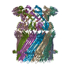
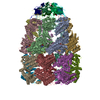
- EMDB-5505: Hexameric structure of the conjugative VirB4 ATPase TrwK: new ins... -
+ Open data

- Basic information
| Entry | Database: EMDB / ID: EMD-5505 | |||||||||
|---|---|---|---|---|---|---|---|---|---|---|
| Title | Hexameric structure of the conjugative VirB4 ATPase TrwK: new insights into a functional and phylogenetic relationship with DNA translocases | |||||||||
 Map data Map data | Reconstruction of hexameric TrwK | |||||||||
 Sample Sample |
| |||||||||
 Keywords Keywords |  bacterial secretion systems / conjugation bacterial secretion systems / conjugation | |||||||||
| Function / homology | CagE, TrbE, VirB component of type IV transporter system / ATP binding Function and homology information Function and homology information | |||||||||
| Biological species |  Escherichia coli (E. coli) Escherichia coli (E. coli) | |||||||||
| Method | single particle reconstruction / negative staining / Resolution: 20.0 Å | |||||||||
 Authors Authors | Pena A / Matilla I / Martin-Benito J / Valpuesta JM / Carrascosa JL / De la Cruz F / Cabezon E / Arechaga I | |||||||||
 Citation Citation | Journal: J Biol Chem / Year: 2012 Title: The hexameric structure of a conjugative VirB4 protein ATPase provides new insights for a functional and phylogenetic relationship with DNA translocases. Authors: Alejandro Peña / Inmaculada Matilla / Jaime Martín-Benito / José M Valpuesta / José L Carrascosa / Fernando de la Cruz / Elena Cabezón / Ignacio Arechaga /  Abstract: VirB4 proteins are ATPases essential for pilus biogenesis and protein transport in type IV secretion systems. These proteins contain a motor domain that shares structural similarities with the motor ...VirB4 proteins are ATPases essential for pilus biogenesis and protein transport in type IV secretion systems. These proteins contain a motor domain that shares structural similarities with the motor domains of DNA translocases, such as the VirD4/TrwB conjugative coupling proteins and the chromosome segregation pump FtsK. Here, we report the three-dimensional structure of full-length TrwK, the VirB4 homologue in the conjugative plasmid R388, determined by single-particle electron microscopy. The structure consists of a hexameric double ring with a barrel-shaped structure. The C-terminal half of VirB4 proteins shares a striking structural similarity with the DNA translocase TrwB. Docking the atomic coordinates of the crystal structures of TrwB and FtsK into the EM map revealed a better fit for FtsK. Interestingly, we have found that like TrwB, TrwK is able to bind DNA with a higher affinity for G4 quadruplex structures than for single-stranded DNA. Furthermore, TrwK exerts a dominant negative effect on the ATPase activity of TrwB, which reflects an interaction between the two proteins. Our studies provide new insights into the structure-function relationship and the evolution of these DNA and protein translocases. | |||||||||
| History |
|
- Structure visualization
| Movie |
Movie viewer |
|---|---|
| Structure viewer | EM map: SurfViewMolmilJmol/JSmol |
| Supplemental images |

- Downloads & links
-EMDB archive
| Map data | emd_5505.map.gz | 571 KB | EMDB map data format | |
|---|---|---|---|---|
| Header (meta data) | emd-5505-v30.xmlemd-5505.xml | 9.6 KB 9.6 KB | Display Display | EMDB header |
| Images | emd_5505.tif | 8.1 MB | ||
| Archive directory |  http://ftp.pdbj.org/pub/emdb/structures/EMD-5505ftp://ftp.pdbj.org/pub/emdb/structures/EMD-5505 http://ftp.pdbj.org/pub/emdb/structures/EMD-5505ftp://ftp.pdbj.org/pub/emdb/structures/EMD-5505 | HTTPS FTP |
-Related structure data
| Similar structure data |
|---|


-Links
| EMDB pages | EMDB (EBI/PDBe) / EMDataResource |
|---|
-Map
| File | Download / File: emd_5505.map.gz / Format: CCP4 / Size: 602.5 KB / Type: IMAGE STORED AS FLOATING POINT NUMBER (4 BYTES) | ||||||||||||||||||||||||||||||||||||||||||||||||||||||||||||||||||||
|---|---|---|---|---|---|---|---|---|---|---|---|---|---|---|---|---|---|---|---|---|---|---|---|---|---|---|---|---|---|---|---|---|---|---|---|---|---|---|---|---|---|---|---|---|---|---|---|---|---|---|---|---|---|---|---|---|---|---|---|---|---|---|---|---|---|---|---|---|---|
| Annotation | Reconstruction of hexameric TrwK | ||||||||||||||||||||||||||||||||||||||||||||||||||||||||||||||||||||
| Voxel size | X=Y=Z: 4.66 Å | ||||||||||||||||||||||||||||||||||||||||||||||||||||||||||||||||||||
| Density |
| ||||||||||||||||||||||||||||||||||||||||||||||||||||||||||||||||||||
| Symmetry | Space group: 1 | ||||||||||||||||||||||||||||||||||||||||||||||||||||||||||||||||||||
| Details | EMDB XML:
CCP4 map header:
| ||||||||||||||||||||||||||||||||||||||||||||||||||||||||||||||||||||
-Supplemental data
- Sample components
-Entire : Hexameric TrwK
| Entire | Name: Hexameric TrwK |
|---|---|
| Components |
|
-Supramolecule #1000: Hexameric TrwK
| Supramolecule | Name: Hexameric TrwK / type: sample / ID: 1000 / Oligomeric state: hexameric / Number unique components: 1 |
|---|---|
| Molecular weight | Theoretical: 540 KDa |
-Macromolecule #1: VirB4 component of type IV transporter system
| Macromolecule | Name: VirB4 component of type IV transporter system / type: protein_or_peptide / ID: 1 / Name.synonym: VirB4 / Oligomeric state: Hexamer / Recombinant expression: Yes |
|---|---|
| Source (natural) | Organism: Escherichia coli (E. coli) |
| Molecular weight | Theoretical: 540 KDa |
| Recombinant expression | Organism: Escherichia coli (E. coli) / Recombinant plasmid: pET |
| Sequence | GO: ATP binding InterPro: CagE, TrbE, VirB component of type IV transporter system |
-Experimental details
-Structure determination
| Method | negative staining |
|---|---|
 Processing Processing | single particle reconstruction |
| Aggregation state | particle |
-Sample preparation
| Concentration | 0.1 mg/mL |
|---|---|
| Buffer | pH: 6.45 Details: 50 mM PIPES-NaOH, 75 mM potassium acetate, 5% (w/v) glycerol, 10 mM magnesium acetate, 0.1 mM EDTA, and 0.5 mM phenylmethylsulfonyl fluoride. |
| Staining | Type: NEGATIVE Details: Samples were negatively stained with 2% (w/v) uranyl acetate |
| Grid | Details: freshly glow-discharged carbon-coated grids |
| Vitrification | Cryogen name: NONE / Instrument: OTHER |
- Electron microscopy
| Microscope | JEOL 1200EXII |
|---|---|
| Electron beam | Acceleration voltage: 100 kV / Electron source: TUNGSTEN HAIRPIN |
| Electron optics | Illumination mode: FLOOD BEAM / Imaging mode: BRIGHT FIELDBright-field microscopy / Cs: 5.6 mm / Nominal defocus max: 1.0 µm / Nominal defocus min: 0.6 µm / Nominal magnification: 60000 |
| Sample stage | Specimen holder model: JEOL |
| Date | Dec 11, 2011 |
| Image recording | Category: FILM / Film or detector model: KODAK SO-163 FILM / Digitization - Scanner: ZEISS SCAI / Average electron dose: 20 e/Å2 |
-Image processing
| CTF correction | Details: Yes |
|---|---|
| Final reconstruction | Algorithm: OTHER / Resolution.type: BY AUTHOR / Resolution: 20.0 Å / Resolution method: FSC 0.5 CUT-OFF / Software - Name: XMIPP / Number images used: 1179 |
