 Movie
Movie Controller


+ Open data

- Basic information
| Entry | Database: EMDB / ID: EMD-5487 | |||||||||
|---|---|---|---|---|---|---|---|---|---|---|
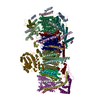
| Title | Rotavirus VP6 protein | |||||||||
 Map data Map data | Reconstruction of rotavirus VP6 coat protein using the DE-12 direct electron detector without movie frame alignment. Icosahedral and 13-fold non-icosahedral averaging was applied. | |||||||||
 Sample Sample |
| |||||||||
 Keywords Keywords | virus coat protein | |||||||||
| Biological species |  Rotavirus Rotavirus | |||||||||
| Method | single particle reconstruction / cryo EM / Resolution: 4.9 Å | |||||||||
 Authors Authors | Campbell MG / Cheng A / Brilot AF / Moeller A / Lyumkis D / Veesler D / Pan J / Harrison SC / Potter CS / Carragher B / Grigorieff N | |||||||||
 Citation Citation | Journal: Structure / Year: 2012 Title: Movies of ice-embedded particles enhance resolution in electron cryo-microscopy. Authors: Melody G Campbell / Anchi Cheng / Axel F Brilot / Arne Moeller / Dmitry Lyumkis / David Veesler / Junhua Pan / Stephen C Harrison / Clinton S Potter / Bridget Carragher / Nikolaus Grigorieff /  Abstract: Low-dose images obtained by electron cryo-microscopy (cryo-EM) are often affected by blurring caused by sample motion during electron beam exposure, degrading signal especially at high resolution. We ...Low-dose images obtained by electron cryo-microscopy (cryo-EM) are often affected by blurring caused by sample motion during electron beam exposure, degrading signal especially at high resolution. We show here that we can align frames of movies, recorded with a direct electron detector during beam exposure of rotavirus double-layered particles, thereby greatly reducing image blurring caused by beam-induced motion and sample stage instabilities. This procedure increases the efficiency of cryo-EM imaging and enhances the resolution obtained in three-dimensional reconstructions of the particle. Using movies in this way is generally applicable to all cryo-EM samples and should also improve the performance of midrange electron microscopes that may have limited mechanical stability and beam coherence. | |||||||||
| History |
|
- Structure visualization
| Movie |
 Movie viewer Movie viewer |
|---|---|
| Structure viewer | EM map: SurfViewMolmilJmol/JSmol |
| Supplemental images |

- Downloads & links
-EMDB archive
| Map data | emd_5487.map.gz | 1.6 MB | EMDB map data format | |
|---|---|---|---|---|
| Header (meta data) | emd-5487-v30.xmlemd-5487.xml | 10.6 KB 10.6 KB | Display Display | EMDB header |
| Images | 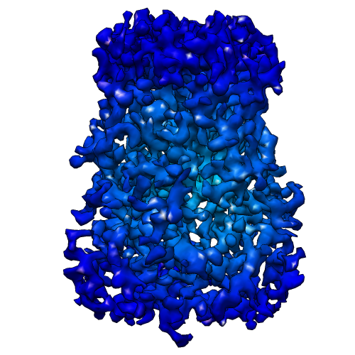 emd_5487_1.png emd_5487_1.png | 195.7 KB | ||
| Archive directory |  http://ftp.pdbj.org/pub/emdb/structures/EMD-5487ftp://ftp.pdbj.org/pub/emdb/structures/EMD-5487 http://ftp.pdbj.org/pub/emdb/structures/EMD-5487ftp://ftp.pdbj.org/pub/emdb/structures/EMD-5487 | HTTPS FTP |
-Validation report
| Summary document | emd_5487_validation.pdf.gz | 78.6 KB | Display | EMDB validaton report |
|---|---|---|---|---|
| Full document | emd_5487_full_validation.pdf.gz | 77.8 KB | Display | |
| Data in XML | emd_5487_validation.xml.gz | 494 B | Display | |
| Arichive directory | https://ftp.pdbj.org/pub/emdb/validation_reports/EMD-5487ftp://ftp.pdbj.org/pub/emdb/validation_reports/EMD-5487 | HTTPS FTP |
-Related structure data


-Links
| EMDB pages | EMDB (EBI/PDBe) / EMDataResource |
|---|
-Map
| File | Download / File: emd_5487.map.gz / Format: CCP4 / Size: 6.5 MB / Type: IMAGE STORED AS FLOATING POINT NUMBER (4 BYTES) | ||||||||||||||||||||||||||||||||||||||||||||||||||||||||||||||||||||
|---|---|---|---|---|---|---|---|---|---|---|---|---|---|---|---|---|---|---|---|---|---|---|---|---|---|---|---|---|---|---|---|---|---|---|---|---|---|---|---|---|---|---|---|---|---|---|---|---|---|---|---|---|---|---|---|---|---|---|---|---|---|---|---|---|---|---|---|---|---|
| Annotation | Reconstruction of rotavirus VP6 coat protein using the DE-12 direct electron detector without movie frame alignment. Icosahedral and 13-fold non-icosahedral averaging was applied. | ||||||||||||||||||||||||||||||||||||||||||||||||||||||||||||||||||||
| Voxel size | X=Y=Z: 1.424 Å | ||||||||||||||||||||||||||||||||||||||||||||||||||||||||||||||||||||
| Density |
| ||||||||||||||||||||||||||||||||||||||||||||||||||||||||||||||||||||
| Symmetry | Space group: 1 | ||||||||||||||||||||||||||||||||||||||||||||||||||||||||||||||||||||
| Details | EMDB XML:
CCP4 map header:
| ||||||||||||||||||||||||||||||||||||||||||||||||||||||||||||||||||||
-Supplemental data
- Sample components
-Entire : Rotavirus VP6 coat protein
| Entire | Name: Rotavirus VP6 coat protein |
|---|---|
| Components |
|
-Supramolecule #1000: Rotavirus VP6 coat protein
| Supramolecule | Name: Rotavirus VP6 coat protein / type: sample / ID: 1000 / Details: The sample was monodisperse / Oligomeric state: Trimer of VP6 / Number unique components: 1 |
|---|---|
| Molecular weight | Theoretical: 41 KDa |
-Macromolecule #1: VP6
| Macromolecule | Name: VP6 / type: protein_or_peptide / ID: 1 / Number of copies: 3 / Oligomeric state: Trimer / Recombinant expression: No / Database: NCBI |
|---|---|
| Source (natural) | Organism: Rotavirus / synonym: Rotavirus |
| Molecular weight | Theoretical: 41 KDa |
-Experimental details
-Structure determination
| Method | cryo EM |
|---|---|
 Processing Processing | single particle reconstruction |
| Aggregation state | particle |
-Sample preparation
| Concentration | 5 mg/mL |
|---|---|
| Buffer | pH: 7.4 |
| Grid | Details: 1.2-1.3 C-flat |
| Vitrification | Cryogen name: ETHANE / Chamber humidity: 85 % / Chamber temperature: 120 K / Instrument: FEI VITROBOT MARK II / Method: Blot for 7 seconds before plunging |
- Electron microscopy
| Microscope | FEI TECNAI F20 |
|---|---|
| Date | Nov 28, 2011 |
| Image recording | Category: CCD / Film or detector model: DIRECT ELECTRON DE-12 (4k x 3k) / Digitization - Sampling interval: 6 µm / Number real images: 501 / Average electron dose: 32 e/Å2 / Details: Movies recorded at 25 frames/second |
| Electron beam | Acceleration voltage: 200 kV / Electron source:  FIELD EMISSION GUN FIELD EMISSION GUN |
| Electron optics | Calibrated magnification: 42135 / Illumination mode: FLOOD BEAM / Imaging mode: BRIGHT FIELD / Cs: 2.0 mm / Nominal defocus max: 2.4 µm / Nominal defocus min: 1.0 µm |
| Sample stage | Specimen holder model: GATAN LIQUID NITROGEN |
| Experimental equipment |  Model: Tecnai F20 / Image courtesy: FEI Company |
-Image processing
| Details | Manual particle selection, refinement using Frealign. |
|---|---|
| CTF correction | Details: Each particle |
| Final reconstruction | Algorithm: OTHER / Resolution.type: BY AUTHOR / Resolution: 4.9 Å / Resolution method: FSC 0.143 CUT-OFF / Software - Name: Frealign, Uppsala_package Details: 13-fold non-icosahedral averaging was applied to the final map Number images used: 807 |
-Atomic model buiding 1
| Initial model | PDB ID: Chain - #0 - Chain ID: C / Chain - #1 - Chain ID: D / Chain - #2 - Chain ID: E |
|---|---|
| Software | Name: Chimera |
| Details | Protocol: Rigid body |
| Refinement | Space: REAL / Protocol: RIGID BODY FIT / Target criteria: Correlation coefficient |

