 Movie
Movie Controller


+ Open data

- Basic information
| Entry | Database: EMDB / ID: EMD-5354 | |||||||||
|---|---|---|---|---|---|---|---|---|---|---|
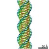
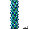
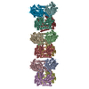
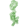
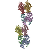
| Title | Remodeling of actin filaments by ADF-cofilin proteins | |||||||||
 Map data Map data | This is the reconstructed volume of the actin-cofilin complex. | |||||||||
 Sample Sample |
| |||||||||
 Keywords Keywords |  actin / cofilin / helical polymers actin / cofilin / helical polymers | |||||||||
| Function / homology |  Function and homology information Function and homology informationGap junction degradation / Formation of annular gap junctions / RHO GTPases activate IQGAPs / UCH proteinases / DNA Damage Recognition in GG-NER / Adherens junctions interactions / Clathrin-mediated endocytosis / RHO GTPases Activate Formins / actin filament fragmentation / positive regulation of embryonic development ...Gap junction degradation / Formation of annular gap junctions / RHO GTPases activate IQGAPs / UCH proteinases / DNA Damage Recognition in GG-NER / Adherens junctions interactions / Clathrin-mediated endocytosis / RHO GTPases Activate Formins / actin filament fragmentation / positive regulation of embryonic development / establishment of spindle localization / structural constituent of postsynaptic actin cytoskeleton / positive regulation by host of viral process / EPH-ephrin mediated repulsion of cells / dense body / actin filament severing / EPHB-mediated forward signaling / VEGFA-VEGFR2 Pathway / regulation of dendritic spine morphogenesis / actin filament depolymerization / RHO GTPases Activate ROCKs / regulation of cell morphogenesis / NuA4 histone acetyltransferase complex / lamellipodium membrane / mitotic cytokinesis / Rho protein signal transduction / Sema3A PAK dependent Axon repulsion / cytoskeleton organization / EPHB-mediated forward signaling / Gene and protein expression by JAK-STAT signaling after Interleukin-12 stimulation / axonogenesis / cell motility / actin filament / response to virus / Regulation of actin dynamics for phagocytic cup formation / ruffle membrane / nuclear matrix / actin filament binding / actin cytoskeleton / Platelet degranulation / lamellipodium / growth cone / actin cytoskeleton organization / vesicle / cytoskeleton / axon / focal adhesion / synapse / negative regulation of apoptotic process / protein kinase binding / extracellular space / extracellular exosome / ATP binding / membrane / nucleus / plasma membrane / cytosol / cytoplasmSimilarity search - Function | |||||||||
| Biological species |  Gallus gallus (chicken) / Gallus gallus (chicken) /  Homo sapiens (human) Homo sapiens (human) | |||||||||
| Method | helical reconstruction / cryo EM / Resolution: 9.0 Å | |||||||||
 Authors Authors | Galkin VE / Orlova A / Kudryashov DS / Solodukhin A / Reisler E / Schoeder GF / Egelman EH | |||||||||
 Citation Citation | Journal: Proc Natl Acad Sci U S A / Year: 2011 Title: Remodeling of actin filaments by ADF/cofilin proteins. Authors: Vitold E Galkin / Albina Orlova / Dmitri S Kudryashov / Alexander Solodukhin / Emil Reisler / Gunnar F Schröder / Edward H Egelman /  Abstract: Cofilin/ADF proteins play key roles in the dynamics of actin, one of the most abundant and highly conserved eukaryotic proteins. We used cryoelectron microscopy to generate a 9-Å resolution three- ...Cofilin/ADF proteins play key roles in the dynamics of actin, one of the most abundant and highly conserved eukaryotic proteins. We used cryoelectron microscopy to generate a 9-Å resolution three-dimensional reconstruction of cofilin-decorated actin filaments, the highest resolution achieved for a complex of F-actin with an actin-binding protein. We show that the cofilin-induced change in the filament twist is due to a unique conformation of the actin molecule unrelated to any previously observed state. The changes between the actin protomer in naked F-actin and in the actin-cofilin filament are greater than the conformational changes between G- and F-actin. Our results show the structural plasticity of actin, suggest that other actin-binding proteins may also induce large but different conformational changes, and show that F-actin cannot be described by a single molecular model. | |||||||||
| History |
|
- Structure visualization
| Movie |
Movie viewer |
|---|---|
| Structure viewer | EM map: SurfViewMolmilJmol/JSmol |
| Supplemental images |

- Downloads & links
-EMDB archive
| Map data | emd_5354.map.gz | 3.6 MB | EMDB map data format | |
|---|---|---|---|---|
| Header (meta data) | emd-5354-v30.xmlemd-5354.xml | 9.6 KB 9.6 KB | Display Display | EMDB header |
| Images | 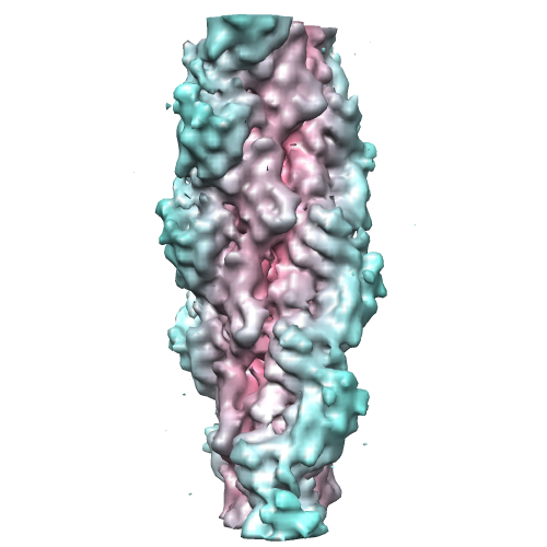 emd_5354_1.png emd_5354_1.png | 141.7 KB | ||
| Archive directory |  http://ftp.pdbj.org/pub/emdb/structures/EMD-5354ftp://ftp.pdbj.org/pub/emdb/structures/EMD-5354 http://ftp.pdbj.org/pub/emdb/structures/EMD-5354ftp://ftp.pdbj.org/pub/emdb/structures/EMD-5354 | HTTPS FTP |
-Related structure data
| Related structure data |  3j0sMC M: atomic model generated by this map C: citing same article ( |
|---|---|
| Similar structure data |
-Links
| EMDB pages | EMDB (EBI/PDBe) / EMDataResource |
|---|---|
| Related items in Molecule of the Month |


-Map
| File | Download / File: emd_5354.map.gz / Format: CCP4 / Size: 3.7 MB / Type: IMAGE STORED AS FLOATING POINT NUMBER (4 BYTES) | ||||||||||||||||||||||||||||||||||||||||||||||||||||||||||||||||||||
|---|---|---|---|---|---|---|---|---|---|---|---|---|---|---|---|---|---|---|---|---|---|---|---|---|---|---|---|---|---|---|---|---|---|---|---|---|---|---|---|---|---|---|---|---|---|---|---|---|---|---|---|---|---|---|---|---|---|---|---|---|---|---|---|---|---|---|---|---|---|
| Annotation | This is the reconstructed volume of the actin-cofilin complex. | ||||||||||||||||||||||||||||||||||||||||||||||||||||||||||||||||||||
| Voxel size | X=Y=Z: 2.5 Å | ||||||||||||||||||||||||||||||||||||||||||||||||||||||||||||||||||||
| Density |
| ||||||||||||||||||||||||||||||||||||||||||||||||||||||||||||||||||||
| Symmetry | Space group: 1 | ||||||||||||||||||||||||||||||||||||||||||||||||||||||||||||||||||||
| Details | EMDB XML:
CCP4 map header:
| ||||||||||||||||||||||||||||||||||||||||||||||||||||||||||||||||||||
-Supplemental data
- Sample components
-Entire : actin decorated with cofilin
| Entire | Name: actin decorated with cofilin |
|---|---|
| Components |
|
-Supramolecule #1000: actin decorated with cofilin
| Supramolecule | Name: actin decorated with cofilin / type: sample / ID: 1000 Oligomeric state: filament containing one cofilin to one actin Number unique components: 2 |
|---|
-Macromolecule #1: F-Actin
| Macromolecule | Name: F-Actin / type: protein_or_peptide / ID: 1 / Name.synonym: F-Actin / Oligomeric state: helical polymer / Recombinant expression: No / Database: NCBI |
|---|---|
| Source (natural) | Organism: Gallus gallus (chicken) / synonym: chicken / Tissue: muscle |
-Macromolecule #2: cofilin-2
| Macromolecule | Name: cofilin-2 / type: protein_or_peptide / ID: 2 / Name.synonym: cofilin-2 / Oligomeric state: one cofilin per actin in filament / Recombinant expression: Yes |
|---|---|
| Source (natural) | Organism: Homo sapiens (human) / synonym: human / Tissue: muscle |
| Recombinant expression | Organism:  Escherichia coli (E. coli) Escherichia coli (E. coli) |
-Experimental details
-Structure determination
| Method | cryo EM |
|---|---|
 Processing Processing | helical reconstruction |
| Aggregation state | filament |
-Sample preparation
| Vitrification | Cryogen name: ETHANE / Chamber humidity: 100 % / Instrument: OTHER / Details: Vitrification instrument: Vitrobot |
|---|
- Electron microscopy
| Microscope | FEI TECNAI F20 |
|---|---|
| Electron beam | Acceleration voltage: 200 kV / Electron source: FIELD EMISSION GUN |
| Electron optics | Illumination mode: FLOOD BEAM / Imaging mode: BRIGHT FIELDBright-field microscopy / Cs: 2.0 mm / Nominal defocus max: 5.3 µm / Nominal defocus min: 1.1 µm / Nominal magnification: 50000 |
| Sample stage | Specimen holder: side entry / Specimen holder model: GATAN LIQUID NITROGEN |
| Date | Jan 1, 2010 |
| Image recording | Category: FILM / Film or detector model: KODAK SO-163 FILM / Digitization - Scanner: NIKON COOLSCAN / Digitization - Sampling interval: 6.35 µm / Number real images: 125 / Bits/pixel: 14 |
| Experimental equipment |  Model: Tecnai F20 / Image courtesy: FEI Company |
-Image processing
| CTF correction | Details: each EM |
|---|---|
| Final reconstruction | Applied symmetry - Helical parameters - Δz: 27.6 Å Applied symmetry - Helical parameters - Δ&Phi: 162.1 ° Algorithm: OTHER / Resolution.type: BY AUTHOR / Resolution: 9.0 Å / Resolution method: OTHER / Software - Name: SPIDER,IHRSR / Details: map calculated from 13,716 segments |
