 Movie
Movie Controller

[English] 日本語
 Yorodumi
- EMDB-5294: 3D reconstruction of frozen hydrated HIV-1 integrase dimer in com... -
+ Open data

- Basic information
| Entry | Database: EMDB / ID: EMD-5294 | |||||||||
|---|---|---|---|---|---|---|---|---|---|---|
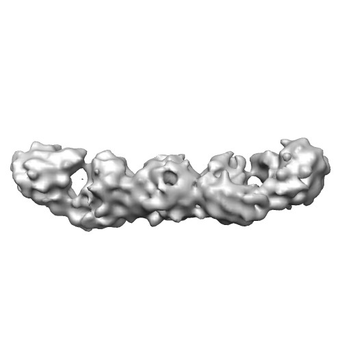
| Title | 3D reconstruction of frozen hydrated HIV-1 integrase dimer in complex with two Fabs. | |||||||||
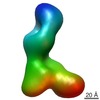
 Map data Map data | This is 3D reconstruction of frozen hydrated HIV-1 integrase dimer in complex with 2 Fabs. | |||||||||
 Sample Sample |
| |||||||||
 Keywords Keywords | HIV-1 integrase dimer /  Fab Fab | |||||||||
| Biological species |   Human immunodeficiency virus 1 Human immunodeficiency virus 1 | |||||||||
| Method | single particle reconstruction / cryo EM / Resolution: 9.6 Å | |||||||||
 Authors Authors | Wu S / Avila-Sakar A / Kim J / Booth DS / Greenberg CH / Rossi A / Liao M / Alian A / Griner SL / Juge N ...Wu S / Avila-Sakar A / Kim J / Booth DS / Greenberg CH / Rossi A / Liao M / Alian A / Griner SL / Juge N / Mergel CM / Chaparro-Riggers J / Strop P / Tampe R / Edwards RH / Stroud RM / Craik CS / Cheng Y | |||||||||
 Citation Citation | Journal: Structure / Year: 2012 Title: Fabs enable single particle cryoEM studies of small proteins. Authors: Shenping Wu / Agustin Avila-Sakar / JungMin Kim / David S Booth / Charles H Greenberg / Andrea Rossi / Maofu Liao / Xueming Li / Akram Alian / Sarah L Griner / Narinobu Juge / Yadong Yu / ...Authors: Shenping Wu / Agustin Avila-Sakar / JungMin Kim / David S Booth / Charles H Greenberg / Andrea Rossi / Maofu Liao / Xueming Li / Akram Alian / Sarah L Griner / Narinobu Juge / Yadong Yu / Claudia M Mergel / Javier Chaparro-Riggers / Pavel Strop / Robert Tampé / Robert H Edwards / Robert M Stroud / Charles S Craik / Yifan Cheng /  Abstract: In spite of its recent achievements, the technique of single particle electron cryomicroscopy (cryoEM) has not been widely used to study proteins smaller than 100 kDa, although it is a highly ...In spite of its recent achievements, the technique of single particle electron cryomicroscopy (cryoEM) has not been widely used to study proteins smaller than 100 kDa, although it is a highly desirable application of this technique. One fundamental limitation is that images of small proteins embedded in vitreous ice do not contain adequate features for accurate image alignment. We describe a general strategy to overcome this limitation by selecting a fragment antigen binding (Fab) to form a stable and rigid complex with a target protein, thus providing a defined feature for accurate image alignment. Using this approach, we determined a three-dimensional structure of an ∼65 kDa protein by single particle cryoEM. Because Fabs can be readily generated against a wide range of proteins by phage display, this approach is generally applicable to study many small proteins by single particle cryoEM. | |||||||||
| History |
|
- Structure visualization
| Movie |
 Movie viewer Movie viewer |
|---|---|
| Structure viewer | EM map: SurfViewMolmilJmol/JSmol |
| Supplemental images |
- Downloads & links
-EMDB archive
| Map data | emd_5294.map.gz | 10.8 MB | EMDB map data format | |
|---|---|---|---|---|
| Header (meta data) | emd-5294-v30.xmlemd-5294.xml | 11.2 KB 11.2 KB | Display Display | EMDB header |
| Images |  emd_5294_1.jpg emd_5294_1.jpg | 20.1 KB | ||
| Archive directory |  http://ftp.pdbj.org/pub/emdb/structures/EMD-5294ftp://ftp.pdbj.org/pub/emdb/structures/EMD-5294 http://ftp.pdbj.org/pub/emdb/structures/EMD-5294ftp://ftp.pdbj.org/pub/emdb/structures/EMD-5294 | HTTPS FTP |
-Related structure data
-Links
| EMDB pages | EMDB (EBI/PDBe) / EMDataResource |
|---|
-Map
| File | Download / File: emd_5294.map.gz / Format: CCP4 / Size: 12.1 MB / Type: IMAGE STORED AS FLOATING POINT NUMBER (4 BYTES) | ||||||||||||||||||||||||||||||||||||||||||||||||||||||||||||||||||||
|---|---|---|---|---|---|---|---|---|---|---|---|---|---|---|---|---|---|---|---|---|---|---|---|---|---|---|---|---|---|---|---|---|---|---|---|---|---|---|---|---|---|---|---|---|---|---|---|---|---|---|---|---|---|---|---|---|---|---|---|---|---|---|---|---|---|---|---|---|---|
| Annotation | This is 3D reconstruction of frozen hydrated HIV-1 integrase dimer in complex with 2 Fabs. | ||||||||||||||||||||||||||||||||||||||||||||||||||||||||||||||||||||
| Voxel size | X=Y=Z: 1.8 Å | ||||||||||||||||||||||||||||||||||||||||||||||||||||||||||||||||||||
| Density |
| ||||||||||||||||||||||||||||||||||||||||||||||||||||||||||||||||||||
| Symmetry | Space group: 1 | ||||||||||||||||||||||||||||||||||||||||||||||||||||||||||||||||||||
| Details | EMDB XML:
CCP4 map header:
| ||||||||||||||||||||||||||||||||||||||||||||||||||||||||||||||||||||
-Supplemental data
- Sample components
-Entire : HIV-1 integrase - Fab complex
| Entire | Name: HIV-1 integrase - Fab complex |
|---|---|
| Components |
|
-Supramolecule #1000: HIV-1 integrase - Fab complex
| Supramolecule | Name: HIV-1 integrase - Fab complex / type: sample / ID: 1000 / Oligomeric state: 2 Fabs bind to one integrase dimer / Number unique components: 2 |
|---|---|
| Molecular weight | Experimental: 160 KDa / Theoretical: 160 KDa |
-Macromolecule #1: integrase
| Macromolecule | Name: integrase / type: protein_or_peptide / ID: 1 / Details: dimer, total molecular weight 65kDa / Number of copies: 2 / Oligomeric state: dimer / Recombinant expression: Yes |
|---|---|
| Source (natural) | Organism: Human immunodeficiency virus 1 / synonym: HIV-1 |
| Molecular weight | Experimental: 32 KDa / Theoretical: 32 KDa |
| Recombinant expression | Organism:  Escherichia coli (E. coli) Escherichia coli (E. coli) |
-Experimental details
-Structure determination
| Method | cryo EM |
|---|---|
 Processing Processing | single particle reconstruction |
| Aggregation state | particle |
-Sample preparation
| Grid | Details: 200 mesh Quantifoil |
|---|---|
| Vitrification | Cryogen name: ETHANE / Chamber humidity: 100 % / Chamber temperature: 100 K / Instrument: OTHER / Details: Vitrification instrument: Vitrobot |
- Electron microscopy
| Microscope | FEI TECNAI F20 |
|---|---|
| Electron beam | Acceleration voltage: 200 kV / Electron source: FIELD EMISSION GUN |
| Electron optics | Illumination mode: FLOOD BEAM / Imaging mode: BRIGHT FIELDBright-field microscopy / Cs: 2.1 mm / Nominal defocus max: 5.0 µm / Nominal defocus min: 2.0 µm / Nominal magnification: 80000 |
| Sample stage | Specimen holder: CT3500 / Specimen holder model: GATAN LIQUID NITROGEN |
| Date | Feb 1, 2011 |
| Image recording | Category: CCD / Film or detector model: TVIPS TEMCAM-F816 (8k x 8k) / Average electron dose: 30 e/Å2 |
| Tilt angle min | 0 |
| Tilt angle max | 0 |
| Experimental equipment |  Model: Tecnai F20 / Image courtesy: FEI Company |
-Image processing
| CTF correction | Details: Each particle |
|---|---|
| Final reconstruction | Algorithm: OTHER / Resolution.type: BY AUTHOR / Resolution: 9.6 Å / Resolution method: FSC 0.143 CUT-OFF / Software - Name: Frealign / Number images used: 14000 |
-Atomic model buiding 1
| Initial model | PDB ID: |
|---|---|
| Software | Name: Chimera |
| Details | Protocol: rigid body |
| Refinement | Space: REAL / Protocol: RIGID BODY FIT |

-Atomic model buiding 2
| Initial model | PDB ID: |
|---|---|
| Software | Name: Chimera |
| Details | Protocol: rigid body |
| Refinement | Space: REAL / Protocol: RIGID BODY FIT |

