 Movie
Movie Controller

[English] 日本語
 Yorodumi
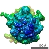
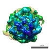
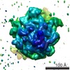
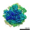
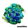
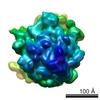
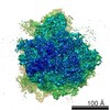
- EMDB-5113: The Structure of Leishmania Mitochondrial Ribosome with Minimal RNA. -
+ Open data

- Basic information
| Entry | Database: EMDB / ID: EMD-5113 | |||||||||
|---|---|---|---|---|---|---|---|---|---|---|
| Title | The Structure of Leishmania Mitochondrial Ribosome with Minimal RNA. | |||||||||
 Map data Map data | Leishmania mitoribosome | |||||||||
 Sample Sample |
| |||||||||
 Keywords Keywords | Leishmania tarentolae / Mitochondrial ribosome / CryoEM / Minimal RNA. | |||||||||
| Function / homology |  Function and homology information Function and homology informationMitochondrial translation elongation / Mitochondrial translation termination / Mitochondrial translation elongation / Mitochondrial translation termination / Mitochondrial translation initiation / mitochondrial large ribosomal subunit / mitochondrial ribosome / mitochondrial translation / misfolded RNA binding / Group I intron splicing ...Mitochondrial translation elongation / Mitochondrial translation termination / Mitochondrial translation elongation / Mitochondrial translation termination / Mitochondrial translation initiation / mitochondrial large ribosomal subunit / mitochondrial ribosome / mitochondrial translation / misfolded RNA binding / Group I intron splicing / RNA folding / four-way junction DNA binding / regulation of mRNA stability / mRNA regulatory element binding translation repressor activity / positive regulation of RNA splicing / DNA endonuclease activity / ribosomal large subunit assembly / maintenance of translational fidelity / mRNA 5'-UTR binding / regulation of translation / ribosomal small subunit assembly / small ribosomal subunit / small ribosomal subunit rRNA binding / large ribosomal subunit rRNA binding / cytosolic small ribosomal subunit / cytosolic large ribosomal subunit / cytoplasmic translation / tRNA binding / molecular adaptor activity / rRNA binding / negative regulation of translation / mitochondrial inner membrane / ribosome / structural constituent of ribosome / translation / protein domain specific binding / response to antibiotic / mRNA binding / mitochondrion / RNA binding / zinc ion binding / nucleoplasm / cytoplasm / cytosol Similarity search - Function | |||||||||
| Biological species |  Leishmania tarentolae (eukaryote) Leishmania tarentolae (eukaryote) | |||||||||
| Method | single particle reconstruction / cryo EM / negative staining / Resolution: 14.1 Å | |||||||||
 Authors Authors | Sharma MR / Booth TM / Simpson L / Maslov DA / Agrawal RK | |||||||||
 Citation Citation | Journal: Proc Natl Acad Sci U S A / Year: 2009 Title: Structure of a mitochondrial ribosome with minimal RNA. Authors: Manjuli R Sharma / Timothy M Booth / Larry Simpson / Dmitri A Maslov / Rajendra K Agrawal /  Abstract: The Leishmania tarentolae mitochondrial ribosome (Lmr) is a minimal ribosomal RNA (rRNA)-containing ribosome. We have obtained a cryo-EM map of the Lmr. The map reveals several features that have not ...The Leishmania tarentolae mitochondrial ribosome (Lmr) is a minimal ribosomal RNA (rRNA)-containing ribosome. We have obtained a cryo-EM map of the Lmr. The map reveals several features that have not been seen in previously-determined structures of eubacterial or eukaryotic (cytoplasmic or organellar) ribosomes to our knowledge. Comparisons of the Lmr map with X-ray crystallographic and cryo-EM maps of the eubacterial ribosomes and a cryo-EM map of the mammalian mitochondrial ribosome show that (i) the overall structure of the Lmr is considerably more porous, (ii) the topology of the intersubunit space is significantly different, with fewer intersubunit bridges, but more tunnels, and (iii) several of the functionally-important rRNA regions, including the alpha-sarcin-ricin loop, have different relative positions within the structure. Furthermore, the major portions of the mRNA channel, the tRNA passage, and the nascent polypeptide exit tunnel contain Lmr-specific proteins, suggesting that the mechanisms for mRNA recruitment, tRNA interaction, and exiting of the nascent polypeptide in Lmr must differ markedly from the mechanisms deduced for ribosomes in other organisms. Our study identifies certain structural features that are characteristic solely of mitochondrial ribosomes and other features that are characteristic of both mitochondrial and chloroplast ribosomes (i.e., organellar ribosomes). | |||||||||
| History |
|
- Structure visualization
| Movie |
Movie viewer |
|---|---|
| Structure viewer | EM map: SurfViewMolmilJmol/JSmol |
| Supplemental images |

- Downloads & links
-EMDB archive
| Map data | emd_5113.map.gz | 7.6 MB | EMDB map data format | |
|---|---|---|---|---|
| Header (meta data) | emd-5113-v30.xmlemd-5113.xml | 9.2 KB 9.2 KB | Display Display | EMDB header |
| Images | 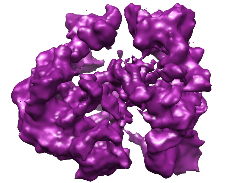 emd_5113_1.jpg emd_5113_1.jpg | 105.9 KB | ||
| Archive directory |  http://ftp.pdbj.org/pub/emdb/structures/EMD-5113ftp://ftp.pdbj.org/pub/emdb/structures/EMD-5113 http://ftp.pdbj.org/pub/emdb/structures/EMD-5113ftp://ftp.pdbj.org/pub/emdb/structures/EMD-5113 | HTTPS FTP |
-Validation report
| Summary document | emd_5113_validation.pdf.gz | 316.3 KB | Display | EMDB validaton report |
|---|---|---|---|---|
| Full document | emd_5113_full_validation.pdf.gz | 315.8 KB | Display | |
| Data in XML | emd_5113_validation.xml.gz | 4.8 KB | Display | |
| Arichive directory | https://ftp.pdbj.org/pub/emdb/validation_reports/EMD-5113ftp://ftp.pdbj.org/pub/emdb/validation_reports/EMD-5113 | HTTPS FTP |
-Related structure data
| Related structure data |  3iy8MC  3iy9MC M: atomic model generated by this map C: citing same article ( |
|---|---|
| Similar structure data |

-Links
| EMDB pages | EMDB (EBI/PDBe) / EMDataResource |
|---|---|
| Related items in Molecule of the Month |


-Map
| File | Download / File: emd_5113.map.gz / Format: CCP4 / Size: 8.2 MB / Type: IMAGE STORED AS FLOATING POINT NUMBER (4 BYTES) | ||||||||||||||||||||||||||||||||||||||||||||||||||||||||||||||||||||
|---|---|---|---|---|---|---|---|---|---|---|---|---|---|---|---|---|---|---|---|---|---|---|---|---|---|---|---|---|---|---|---|---|---|---|---|---|---|---|---|---|---|---|---|---|---|---|---|---|---|---|---|---|---|---|---|---|---|---|---|---|---|---|---|---|---|---|---|---|---|
| Annotation | Leishmania mitoribosome | ||||||||||||||||||||||||||||||||||||||||||||||||||||||||||||||||||||
| Voxel size | X=Y=Z: 2.76 Å | ||||||||||||||||||||||||||||||||||||||||||||||||||||||||||||||||||||
| Density |
| ||||||||||||||||||||||||||||||||||||||||||||||||||||||||||||||||||||
| Symmetry | Space group: 1 | ||||||||||||||||||||||||||||||||||||||||||||||||||||||||||||||||||||
| Details | EMDB XML:
CCP4 map header:
| ||||||||||||||||||||||||||||||||||||||||||||||||||||||||||||||||||||
-Supplemental data
- Sample components
-Entire : Leishmania Mitochondrial 50S Ribosome
| Entire | Name: Leishmania Mitochondrial 50S Ribosome |
|---|---|
| Components |
|
-Supramolecule #1000: Leishmania Mitochondrial 50S Ribosome
| Supramolecule | Name: Leishmania Mitochondrial 50S Ribosome / type: sample / ID: 1000 / Oligomeric state: Monomer / Number unique components: 1 |
|---|---|
| Molecular weight | Experimental: 2.1 MDa / Method: Proteomics |
-Supramolecule #1: 50S Ribosome
| Supramolecule | Name: 50S Ribosome / type: complex / ID: 1 / Recombinant expression: No / Database: NCBI / Ribosome-details: ribosome-prokaryote: LSU 50S |
|---|---|
| Source (natural) | Organism: Leishmania tarentolae (eukaryote) / Organelle: mitochondrion |
| Molecular weight | Experimental: 2.1 MDa |
-Experimental details
-Structure determination
| Method | negative staining, cryo EM |
|---|---|
 Processing Processing | single particle reconstruction |
| Aggregation state | particle |
-Sample preparation
| Concentration | 0.067 mg/mL |
|---|---|
| Buffer | pH: 7.5 Details: 50 mM Tris-HCl, pH 7.5, 100 mM KCl, 10 mM MgCl2, 3 mM DTT, 0.1 mM EDTA and 0.05% dodecyl maltoside |
| Staining | Type: NEGATIVE / Details: Cryo EM |
| Grid | Details: 300 copper Mesh grid |
| Vitrification | Cryogen name: ETHANE / Chamber humidity: 90 % / Instrument: OTHER |
- Electron microscopy
| Microscope | FEI TECNAI F20 |
|---|---|
| Image recording | Category: FILM / Film or detector model: KODAK SO-163 FILM / Digitization - Scanner: ZEISS SCAI / Digitization - Sampling interval: 14 µm / Number real images: 267 / Bits/pixel: 12 |
| Electron beam | Acceleration voltage: 200 kV / Electron source:  FIELD EMISSION GUN FIELD EMISSION GUN |
| Electron optics | Calibrated magnification: 50760 / Illumination mode: FLOOD BEAM / Imaging mode: BRIGHT FIELD / Nominal defocus max: 4.5 µm / Nominal defocus min: 1.6 µm / Nominal magnification: 50000 |
| Sample stage | Specimen holder: eucentric / Specimen holder model: OTHER |
| Experimental equipment |  Model: Tecnai F20 / Image courtesy: FEI Company |
-Image processing
| CTF correction | Details: Each Micrograph |
|---|---|
| Final reconstruction | Algorithm: OTHER / Resolution.type: BY AUTHOR / Resolution: 14.1 Å / Resolution method: FSC 0.5 CUT-OFF / Software - Name: SPIDER / Number images used: 53475 |
