 Movie
Movie Controller

[English] 日本語
 Yorodumi
- EMDB-3384: A closed conformation of the C. elegans separase-securin complex -
+ Open data

- Basic information
| Entry | Database: EMDB / ID: EMD-3384 | |||||||||
|---|---|---|---|---|---|---|---|---|---|---|
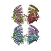
| Title | A closed conformation of the C. elegans separase-securin complex | |||||||||
 Map data Map data | Reconstruction of C. elegans separase-securinn complex | |||||||||
 Sample Sample |
| |||||||||
 Keywords Keywords |  chromosome segregation / protease chromosome segregation / protease | |||||||||
| Function / homology |  Function and homology information Function and homology informationSeparation of Sister Chromatids / separase-securin complex / eggshell formation / metaphase plate / regulation of nematode larval development / separase / cortical granule exocytosis / maintenance of meiotic sister chromatid cohesion / meiotic chromosome separation / maintenance of mitotic sister chromatid cohesion ...Separation of Sister Chromatids / separase-securin complex / eggshell formation / metaphase plate / regulation of nematode larval development / separase / cortical granule exocytosis / maintenance of meiotic sister chromatid cohesion / meiotic chromosome separation / maintenance of mitotic sister chromatid cohesion / polarity specification of anterior/posterior axis / polar body extrusion after meiotic divisions / cortical granule / regulation of centriole-centriole cohesion / mitotic sister chromatid separation / regulation of exocytosis / multicellular organismal reproductive process / meiotic spindle / regulation of locomotion / centrosome localization / cleavage furrow / centrosome duplication / mitotic cytokinesis / condensed chromosome / condensed nuclear chromosome / spindle microtubule / protein localization / spindle / mitotic spindle / protein processing / nuclear envelope / chromosome / cell cortex / midbody / protease binding / protein stabilization / cysteine-type endopeptidase activity / centrosome / ubiquitin protein ligase binding / nucleus / cytoplasmSimilarity search - Function | |||||||||
| Biological species |  Caenorhabditis elegans (invertebrata) Caenorhabditis elegans (invertebrata) | |||||||||
| Method | single particle reconstruction / negative staining / Resolution: 24.0 Å | |||||||||
 Authors Authors | Bachmann G / Morris E / Bayliss R | |||||||||
 Citation Citation | Journal: Open Biol / Year: 2016 Title: A closed conformation of the Caenorhabditis elegans separase-securin complex. Authors: Gudrun Bachmann / Mark W Richards / Anja Winter / Fabienne Beuron / Edward Morris / Richard Bayliss /  Abstract: The protease separase plays a key role in sister chromatid disjunction and centriole disengagement. To maintain genomic stability, separase activity is strictly regulated by binding of an inhibitory ...The protease separase plays a key role in sister chromatid disjunction and centriole disengagement. To maintain genomic stability, separase activity is strictly regulated by binding of an inhibitory protein, securin. Despite its central role in cell division, the separase and securin complex is poorly understood at the structural level. This is partly owing to the difficulty of generating a sufficient quantity of homogeneous, stable protein. Here, we report the production of Caenorhabditis elegans separase-securin complex, and its characterization using biochemical methods and by negative staining electron microscopy. Single particle analysis generated a density map at a resolution of 21-24 Å that reveals a close, globular structure of complex connectivity harbouring two lobes. One lobe matches closely a homology model of the N-terminal HEAT repeat domain of separase, whereas the second lobe readily accommodates homology models of the separase C-terminal death and caspase-like domains. The globular structure of the C. elegans separase-securin complex contrasts with the more elongated structure previously described for the Homo sapiens complex, which could represent a different functional state of the complex, suggesting a mechanism for the regulation of separase activity through conformational change. | |||||||||
| History |
|
- Structure visualization
| Movie |
Movie viewer |
|---|---|
| Structure viewer | EM map: SurfViewMolmilJmol/JSmol |
| Supplemental images |

- Downloads & links
-EMDB archive
| Map data | emd_3384.map.gz | 3.6 MB | EMDB map data format | |
|---|---|---|---|---|
| Header (meta data) | emd-3384-v30.xmlemd-3384.xml | 9.3 KB 9.3 KB | Display Display | EMDB header |
| Images | 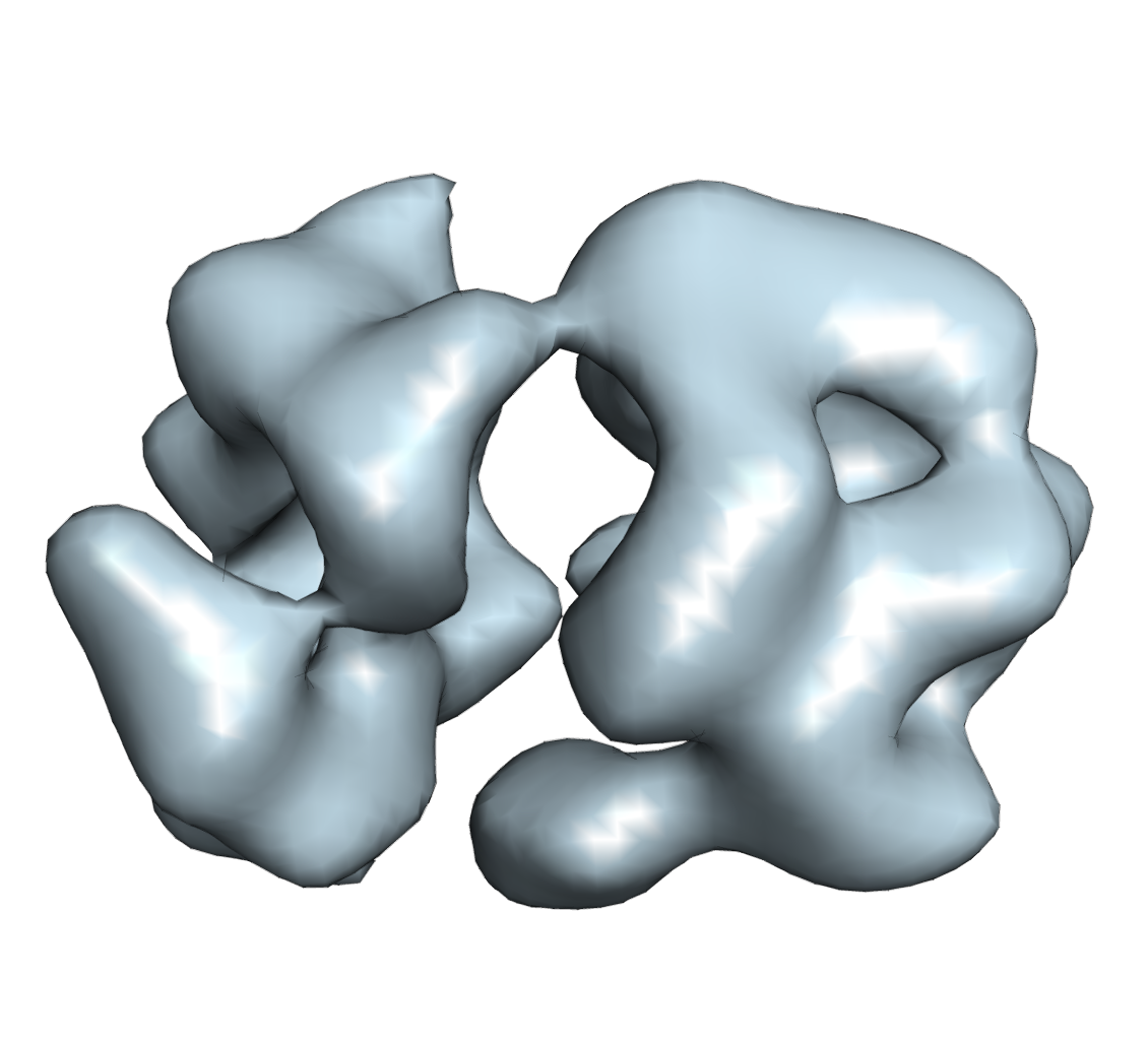 3384.png 3384.png | 354.1 KB | ||
| Archive directory |  http://ftp.pdbj.org/pub/emdb/structures/EMD-3384ftp://ftp.pdbj.org/pub/emdb/structures/EMD-3384 http://ftp.pdbj.org/pub/emdb/structures/EMD-3384ftp://ftp.pdbj.org/pub/emdb/structures/EMD-3384 | HTTPS FTP |
-Related structure data
| Similar structure data |
|---|


-Links
| EMDB pages | EMDB (EBI/PDBe) / EMDataResource |
|---|
-Map
| File | Download / File: emd_3384.map.gz / Format: CCP4 / Size: 3.7 MB / Type: IMAGE STORED AS FLOATING POINT NUMBER (4 BYTES) | ||||||||||||||||||||||||||||||||||||||||||||||||||||||||||||
|---|---|---|---|---|---|---|---|---|---|---|---|---|---|---|---|---|---|---|---|---|---|---|---|---|---|---|---|---|---|---|---|---|---|---|---|---|---|---|---|---|---|---|---|---|---|---|---|---|---|---|---|---|---|---|---|---|---|---|---|---|---|
| Annotation | Reconstruction of C. elegans separase-securinn complex | ||||||||||||||||||||||||||||||||||||||||||||||||||||||||||||
| Voxel size | X=Y=Z: 2.915 Å | ||||||||||||||||||||||||||||||||||||||||||||||||||||||||||||
| Density |
| ||||||||||||||||||||||||||||||||||||||||||||||||||||||||||||
| Symmetry | Space group: 1 | ||||||||||||||||||||||||||||||||||||||||||||||||||||||||||||
| Details | EMDB XML:
CCP4 map header:
| ||||||||||||||||||||||||||||||||||||||||||||||||||||||||||||
-Supplemental data
- Sample components
-Entire : separase-securin complex
| Entire | Name: separase-securin complex |
|---|---|
| Components |
|
-Supramolecule #1000: separase-securin complex
| Supramolecule | Name: separase-securin complex / type: sample / ID: 1000 / Details: The sample was monodisperse Oligomeric state: one monomer of separase bound to one monomer of securin Number unique components: 2 |
|---|---|
| Molecular weight | Experimental: 174 KDa / Theoretical: 171 KDa / Method: Multi-angle light scattering |
-Macromolecule #1: separase
| Macromolecule | Name: separase / type: protein_or_peptide / ID: 1 / Number of copies: 1 / Oligomeric state: momomer / Recombinant expression: Yes |
|---|---|
| Source (natural) | Organism: Caenorhabditis elegans (invertebrata) |
| Molecular weight | Theoretical: 144 KDa |
| Recombinant expression | Organism:   Spodoptera frugiperda (fall armyworm) Spodoptera frugiperda (fall armyworm) |
| Sequence | UniProtKB: Separin homolog sep-1 |
-Macromolecule #2: securin
| Macromolecule | Name: securin / type: protein_or_peptide / ID: 2 / Number of copies: 1 / Oligomeric state: monomer / Recombinant expression: Yes |
|---|---|
| Source (natural) | Organism: Caenorhabditis elegans (invertebrata) |
| Molecular weight | Theoretical: 27 KDa |
| Recombinant expression | Organism: Spodoptera frugiperda (fall armyworm) |
| Sequence | UniProtKB: Securin-like protein |
-Experimental details
-Structure determination
| Method | negative staining |
|---|---|
 Processing Processing | single particle reconstruction |
| Aggregation state | particle |
-Sample preparation
| Concentration | 0.1 mg/mL |
|---|---|
| Buffer | pH: 7 / Details: 50mM Tris, 0.5M NaCl, 2mM beta-mercaptoethanol |
| Staining | Type: NEGATIVE Details: Grids with adsorbed protein floated on 1% w/v uranyl acetate for 20 seconds. |
| Grid | Details: Thin carbon support on Quantifoil R 1.2/1.3 grids glow discharged in residual air |
| Vitrification | Cryogen name: NONE / Instrument: OTHER |
- Electron microscopy
| Microscope | FEI TECNAI F20 |
|---|---|
| Electron beam | Acceleration voltage: 200 kV / Electron source: FIELD EMISSION GUN |
| Electron optics | Illumination mode: FLOOD BEAM / Imaging mode: BRIGHT FIELDBright-field microscopy / Cs: 2.0 mm / Nominal defocus max: 0.85 µm / Nominal defocus min: 0.7 µm / Nominal magnification: 62000 |
| Sample stage | Specimen holder model: SIDE ENTRY, EUCENTRIC |
| Date | Nov 10, 2010 |
| Image recording | Category: CCD / Film or detector model: TVIPS TEMCAM-F415 (4k x 4k) / Digitization - Sampling interval: 15 µm / Average electron dose: 100 e/Å2 / Bits/pixel: 16 |
| Experimental equipment |  Model: Tecnai F20 / Image courtesy: FEI Company |
-Image processing
| Final reconstruction | Applied symmetry - Point group: C1 (asymmetric) / Algorithm: OTHER / Resolution.type: BY AUTHOR / Resolution: 24.0 Å / Resolution method: OTHER / Software - Name: Imagic, Spider / Number images used: 5764 |
|---|
