 Movie
Movie Controller

[English] 日本語
 Yorodumi
- EMDB-3322: Multiple capsid-stabilizing interactions revealed in a high-resol... -
+ Open data

- Basic information
| Entry | Database: EMDB / ID: EMD-3322 | |||||||||
|---|---|---|---|---|---|---|---|---|---|---|
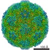
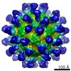
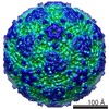
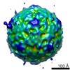
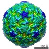
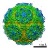
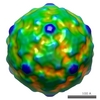
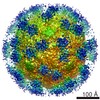
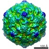
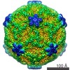
| Title | Multiple capsid-stabilizing interactions revealed in a high-resolution structure of an emerging picornavirus causing neonatal sepsis | |||||||||
 Map data Map data | Asymmetric reconstruction of human parechovirus 3 (HPeV3). | |||||||||
 Sample Sample |
| |||||||||
 Keywords Keywords | asymmetric reconstruction / HPeV3 / parechovirus / picornavirus / single particle anaylsis / human parechovirus / neonatal sepsis / cryoEM / image processing | |||||||||
| Biological species |  Human parechovirus 3 Human parechovirus 3 | |||||||||
| Method | single particle reconstruction / cryo EM / negative staining / Resolution: 10.36 Å | |||||||||
 Authors Authors | Shakeel S / Westerhuis BM / Domanska A / Koning RI / Matadeen R / Koster AJ / Bakker AQ / Beaumont T / Wolthers KC / Butcher SJ | |||||||||
 Citation Citation | Journal: Nat Commun / Year: 2016 Title: Multiple capsid-stabilizing interactions revealed in a high-resolution structure of an emerging picornavirus causing neonatal sepsis. Authors: Shabih Shakeel / Brenda M Westerhuis / Ausra Domanska / Roman I Koning / Rishi Matadeen / Abraham J Koster / Arjen Q Bakker / Tim Beaumont / Katja C Wolthers / Sarah J Butcher /   Abstract: The poorly studied picornavirus, human parechovirus 3 (HPeV3) causes neonatal sepsis with no therapies available. Our 4.3-Å resolution structure of HPeV3 on its own and at 15 Å resolution in ...The poorly studied picornavirus, human parechovirus 3 (HPeV3) causes neonatal sepsis with no therapies available. Our 4.3-Å resolution structure of HPeV3 on its own and at 15 Å resolution in complex with human monoclonal antibody Fabs demonstrates the expected picornavirus capsid structure with three distinct features. First, 25% of the HPeV3 RNA genome in 60 sites is highly ordered as confirmed by asymmetric reconstruction, and interacts with conserved regions of the capsid proteins VP1 and VP3. Second, the VP0 N terminus stabilizes the capsid inner surface, in contrast to other picornaviruses where on expulsion as VP4, it forms an RNA translocation channel. Last, VP1's hydrophobic pocket, the binding site for the antipicornaviral drug, pleconaril, is blocked and thus inappropriate for antiviral development. Together, these results suggest a direction for development of neutralizing antibodies, antiviral drugs based on targeting the RNA-protein interactions and dissection of virus assembly on the basis of RNA nucleation. | |||||||||
| History |
|
- Structure visualization
| Movie |
 Movie viewer Movie viewer |
|---|---|
| Structure viewer | EM map: SurfViewMolmilJmol/JSmol |
| Supplemental images |

- Downloads & links
-EMDB archive
| Map data | emd_3322.map.gz | 190.4 MB | EMDB map data format | |
|---|---|---|---|---|
| Header (meta data) | emd-3322-v30.xmlemd-3322.xml | 10.1 KB 10.1 KB | Display Display | EMDB header |
| FSC (resolution estimation) | emd_3322_fsc.xml | 16.7 KB | Display | FSC data file |
| Images | emd_3322.tif | 71.6 KB | ||
| Archive directory |  http://ftp.pdbj.org/pub/emdb/structures/EMD-3322ftp://ftp.pdbj.org/pub/emdb/structures/EMD-3322 http://ftp.pdbj.org/pub/emdb/structures/EMD-3322ftp://ftp.pdbj.org/pub/emdb/structures/EMD-3322 | HTTPS FTP |
-Validation report
| Summary document | emd_3322_validation.pdf.gz | 261.3 KB | Display | EMDB validaton report |
|---|---|---|---|---|
| Full document | emd_3322_full_validation.pdf.gz | 260.5 KB | Display | |
| Data in XML | emd_3322_validation.xml.gz | 14 KB | Display | |
| Arichive directory | https://ftp.pdbj.org/pub/emdb/validation_reports/EMD-3322ftp://ftp.pdbj.org/pub/emdb/validation_reports/EMD-3322 | HTTPS FTP |
-Related structure data

-Links
| EMDB pages | EMDB (EBI/PDBe) / EMDataResource |
|---|
-Map
| File | Download / File: emd_3322.map.gz / Format: CCP4 / Size: 238.4 MB / Type: IMAGE STORED AS FLOATING POINT NUMBER (4 BYTES) | ||||||||||||||||||||||||||||||||||||||||||||||||||||||||||||
|---|---|---|---|---|---|---|---|---|---|---|---|---|---|---|---|---|---|---|---|---|---|---|---|---|---|---|---|---|---|---|---|---|---|---|---|---|---|---|---|---|---|---|---|---|---|---|---|---|---|---|---|---|---|---|---|---|---|---|---|---|---|
| Annotation | Asymmetric reconstruction of human parechovirus 3 (HPeV3). | ||||||||||||||||||||||||||||||||||||||||||||||||||||||||||||
| Voxel size | X=Y=Z: 1.14 Å | ||||||||||||||||||||||||||||||||||||||||||||||||||||||||||||
| Density |
| ||||||||||||||||||||||||||||||||||||||||||||||||||||||||||||
| Symmetry | Space group: 1 | ||||||||||||||||||||||||||||||||||||||||||||||||||||||||||||
| Details | EMDB XML:
CCP4 map header:
| ||||||||||||||||||||||||||||||||||||||||||||||||||||||||||||
-Supplemental data
- Sample components
-Entire : Human parechovirus 3 virions
| Entire | Name: Human parechovirus 3 virions |
|---|---|
| Components |
|
-Supramolecule #1000: Human parechovirus 3 virions
| Supramolecule | Name: Human parechovirus 3 virions / type: sample / ID: 1000 / Details: The sample was monodisperse / Number unique components: 1 |
|---|
-Supramolecule #1: Human parechovirus 3
| Supramolecule | Name: Human parechovirus 3 / type: virus / ID: 1 / Name.synonym: HPeV3 / NCBI-ID: 195055 / Sci species name: Human parechovirus 3 / Virus type: VIRION / Virus isolate: OTHER / Virus enveloped: No / Virus empty: No / Syn species name: HPeV3 |
|---|---|
| Host (natural) | Organism:  Homo sapiens (human) / synonym: VERTEBRATES Homo sapiens (human) / synonym: VERTEBRATES |
| Host system | Organism:  Chlorocebus aethiops (grivet) / Recombinant strain: African green monkey / Recombinant cell: Vero Chlorocebus aethiops (grivet) / Recombinant strain: African green monkey / Recombinant cell: Vero |
| Virus shell | Shell ID: 1 / Diameter: 280 Å |
-Experimental details
-Structure determination
| Method | negative staining, cryo EM |
|---|---|
 Processing Processing | single particle reconstruction |
| Aggregation state | particle |
-Sample preparation
| Concentration | 1 mg/mL |
|---|---|
| Buffer | pH: 7.5 / Details: 10mM Tris-HCl, 150mM NaCl, 1mM MgCl2 |
| Staining | Type: NEGATIVE / Details: unstained sample |
| Grid | Details: Quantifoil holey carbon on copper grids |
| Vitrification | Cryogen name: ETHANE / Instrument: LEICA EM GP / Method: Blot for 2 sec on one side before plunging. |
- Electron microscopy
| Microscope | FEI TITAN KRIOS |
|---|---|
| Temperature | Min: 80 K / Max: 95 K / Average: 87 K |
| Alignment procedure | Legacy - Astigmatism: Done as part of Cs corector routine. |
| Details | Cs corrector was used during imaging. |
| Date | Jan 21, 2014 |
| Image recording | Category: CCD / Film or detector model: FEI FALCON II (4k x 4k) / Number real images: 6604 / Average electron dose: 36 e/Å2 Details: Total number of images used in the reconstruction were 1028. |
| Electron beam | Acceleration voltage: 300 kV / Electron source:  FIELD EMISSION GUN FIELD EMISSION GUN |
| Electron optics | Illumination mode: FLOOD BEAM / Imaging mode: BRIGHT FIELD / Cs: 0.01 mm / Nominal defocus max: 2.34 µm / Nominal defocus min: 0.42 µm / Nominal magnification: 59000 |
| Sample stage | Specimen holder: Liquid nitrogen cooled. / Specimen holder model: FEI TITAN KRIOS AUTOGRID HOLDER |
| Experimental equipment |  Model: Titan Krios / Image courtesy: FEI Company |
-Image processing
| Details | The movie frames were aligned initially by motion_corr. CTF deteremined using CTFFIND3. Particles picked using Ethan. 2D and 3D classification (with icosahedral symmetry imposed) done in Relion. Final reconstruction in Relion. |
|---|---|
| CTF correction | Details: Each micrograph |
| Final reconstruction | Applied symmetry - Point group: C1 (asymmetric) / Algorithm: OTHER / Resolution.type: BY AUTHOR / Resolution: 10.36 Å / Resolution method: OTHER Software - Name: Ethan, CTFFIND3, Eman1, Eman2, Auto3DEM, Relion, ResMap Number images used: 41845 |
| FSC plot (resolution estimation) |  |
