 Movie
Movie Controller

[English] 日本語
 Yorodumi
- EMDB-3197: Sub-tomogram averaging of electron cryo-microscopic data taken fr... -
+ Open data

- Basic information
| Entry | Database: EMDB / ID: EMD-3197 | |||||||||
|---|---|---|---|---|---|---|---|---|---|---|
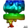
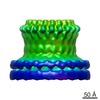
| Title | Sub-tomogram averaging of electron cryo-microscopic data taken from focused-ion beam milled lamellae of nuclei of Pseudorabies virus (PrV) nuclear egress complex-expressing cells | |||||||||
 Map data Map data | Sub-tomogram average of nuclear egress complex from vesicles observed inside nuclei of cells co-expressing PrV UL31 and UL34 by focus-ion beam milling and electron cryo-tomography. | |||||||||
 Sample Sample |
| |||||||||
 Keywords Keywords |  alphaherpesvirinae / herpesvirus simplex / HSV-1 / pseudorabies virus / PrV / nuclear egress complex / nuclear envelope / nucleoplasmic reticulum / inner nuclear membrane / UL31 / UL34 / vesicle transport / nucleo-cytoplasmic transport / nanovesicles / cryoEM / cryoET / electron cryo-microscopy / electron cryo-tomography / cryoFIB / focused ion beam milling / FIB-SEM alphaherpesvirinae / herpesvirus simplex / HSV-1 / pseudorabies virus / PrV / nuclear egress complex / nuclear envelope / nucleoplasmic reticulum / inner nuclear membrane / UL31 / UL34 / vesicle transport / nucleo-cytoplasmic transport / nanovesicles / cryoEM / cryoET / electron cryo-microscopy / electron cryo-tomography / cryoFIB / focused ion beam milling / FIB-SEM | |||||||||
| Function / homology |  Function and homology information Function and homology informationhost cell nuclear inner membrane / viral budding from nuclear membrane / membrane / metal ion bindingSimilarity search - Function | |||||||||
| Biological species |  Sus scrofa (pig) / Sus scrofa (pig) /  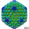 Suid herpesvirus 1 Suid herpesvirus 1 | |||||||||
| Method | subtomogram averaging / cryo EM / negative staining / Resolution: 35.0 Å | |||||||||
 Authors Authors | Hagen C / Siebert CA / Dent KC / Vasishtan D / Zeev Ben Mordehai T / Grange M / Klupp BG / Mettenleiter T / Gruenewald K | |||||||||
 Citation Citation | Journal: Cell / Year: 2015 Title: Structural Basis of Vesicle Formation at the Inner Nuclear Membrane. Authors: Christoph Hagen / Kyle C Dent / Tzviya Zeev-Ben-Mordehai / Michael Grange / Jens B Bosse / Cathy Whittle / Barbara G Klupp / C Alistair Siebert / Daven Vasishtan / Felix J B Bäuerlein / ...Authors: Christoph Hagen / Kyle C Dent / Tzviya Zeev-Ben-Mordehai / Michael Grange / Jens B Bosse / Cathy Whittle / Barbara G Klupp / C Alistair Siebert / Daven Vasishtan / Felix J B Bäuerlein / Juliana Cheleski / Stephan Werner / Peter Guttmann / Stefan Rehbein / Katja Henzler / Justin Demmerle / Barbara Adler / Ulrich Koszinowski / Lothar Schermelleh / Gerd Schneider / Lynn W Enquist / Jürgen M Plitzko / Thomas C Mettenleiter / Kay Grünewald /    Abstract: Vesicular nucleo-cytoplasmic transport is becoming recognized as a general cellular mechanism for translocation of large cargoes across the nuclear envelope. Cargo is recruited, enveloped at the ...Vesicular nucleo-cytoplasmic transport is becoming recognized as a general cellular mechanism for translocation of large cargoes across the nuclear envelope. Cargo is recruited, enveloped at the inner nuclear membrane (INM), and delivered by membrane fusion at the outer nuclear membrane. To understand the structural underpinning for this trafficking, we investigated nuclear egress of progeny herpesvirus capsids where capsid envelopment is mediated by two viral proteins, forming the nuclear egress complex (NEC). Using a multi-modal imaging approach, we visualized the NEC in situ forming coated vesicles of defined size. Cellular electron cryo-tomography revealed a protein layer showing two distinct hexagonal lattices at its membrane-proximal and membrane-distant faces, respectively. NEC coat architecture was determined by combining this information with integrative modeling using small-angle X-ray scattering data. The molecular arrangement of the NEC establishes the basic mechanism for budding and scission of tailored vesicles at the INM. #1: Journal: Cell Reports / Year: 2015Title: Crystal structure of the herpesvirus nuclear egress complex provides insights into inner nuclear membrane remodelling Authors: Zeev-Ben-Mordehai T / Weberruss M / Lorenz M / Cheleski J / Hellberg T / Whittle C / El Omari K / Vasishtan D / Dent KC Harlos K / Franzke K / Hagen C / Klupp B / Antonin W / Mettenleiter TC / Gruenewald K | |||||||||
| History |
|
- Structure visualization
| Movie |
Movie viewer |
|---|---|
| Structure viewer | EM map: SurfViewMolmilJmol/JSmol |
| Supplemental images |

- Downloads & links
-EMDB archive
| Map data | emd_3197.map.gz | 28.4 KB | EMDB map data format | |
|---|---|---|---|---|
| Header (meta data) | emd-3197-v30.xmlemd-3197.xml | 15.5 KB 15.5 KB | Display Display | EMDB header |
| Images |  emd_3197.png emd_3197.png | 51 KB | ||
| Archive directory |  http://ftp.pdbj.org/pub/emdb/structures/EMD-3197ftp://ftp.pdbj.org/pub/emdb/structures/EMD-3197 http://ftp.pdbj.org/pub/emdb/structures/EMD-3197ftp://ftp.pdbj.org/pub/emdb/structures/EMD-3197 | HTTPS FTP |
-Related structure data
| Related structure data |  5fkiM 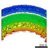 3215C M: atomic model generated by this map C: citing same article ( |
|---|---|
| Similar structure data |
-Links
| EMDB pages | EMDB (EBI/PDBe) / EMDataResource |
|---|
-Map
| File | Download / File: emd_3197.map.gz / Format: CCP4 / Size: 32.2 KB / Type: IMAGE STORED AS FLOATING POINT NUMBER (4 BYTES) | ||||||||||||||||||||||||||||||||||||||||||||||||||||||||||||
|---|---|---|---|---|---|---|---|---|---|---|---|---|---|---|---|---|---|---|---|---|---|---|---|---|---|---|---|---|---|---|---|---|---|---|---|---|---|---|---|---|---|---|---|---|---|---|---|---|---|---|---|---|---|---|---|---|---|---|---|---|---|
| Annotation | Sub-tomogram average of nuclear egress complex from vesicles observed inside nuclei of cells co-expressing PrV UL31 and UL34 by focus-ion beam milling and electron cryo-tomography. | ||||||||||||||||||||||||||||||||||||||||||||||||||||||||||||
| Voxel size | X=Y=Z: 11.4 Å | ||||||||||||||||||||||||||||||||||||||||||||||||||||||||||||
| Density |
| ||||||||||||||||||||||||||||||||||||||||||||||||||||||||||||
| Symmetry | Space group: 1 | ||||||||||||||||||||||||||||||||||||||||||||||||||||||||||||
| Details | EMDB XML:
CCP4 map header:
| ||||||||||||||||||||||||||||||||||||||||||||||||||||||||||||
-Supplemental data
- Sample components
-Entire : vesicles coated with pUL31/pUL34 heterodimers
| Entire | Name: vesicles coated with pUL31/pUL34 heterodimers |
|---|---|
| Components |
|
-Supramolecule #1000: vesicles coated with pUL31/pUL34 heterodimers
| Supramolecule | Name: vesicles coated with pUL31/pUL34 heterodimers / type: sample / ID: 1000 Details: porcine epithelial-like embryonic EFN-R kidney cells stably co-expressing PrV UL31 and UL34, the latter fused with GFP (cell line designated as BK/EF/UL31/34gfp catalogue No. RIE 1083 of the ...Details: porcine epithelial-like embryonic EFN-R kidney cells stably co-expressing PrV UL31 and UL34, the latter fused with GFP (cell line designated as BK/EF/UL31/34gfp catalogue No. RIE 1083 of the Collection of Cell Lines in Veterinary Medicine at the FLI, Greifswald-Insel Riems, Germany) Oligomeric state: coat of ~500 hexamers of pUL31/pUL34 heterodimers per vesicle Number unique components: 2 |
|---|
-Supramolecule #1: Nuclear envelope
| Supramolecule | Name: Nuclear envelope / type: organelle_or_cellular_component / ID: 1 / Name.synonym: Inner and outer nuclear membranes Details: two proteins (pUL31 and pUL34) of pseudorabies virus (PrV) are co-expressed in the cells forming there the herpesviral nuclear egress complex lining as a coat perinuclear vesicles Oligomeric state: heterodimer / Recombinant expression: No |
|---|---|
| Ref INTERPRO | divclassse qspanoncli ckpopupspa nclassgree n(this)spandata popltspanc lassquotlo adingbarqu otgtltimgs rcquotimgl oadinggifq uotdecodin gquotasync quotgtltsp angtdataur lajaxphp?m odetaxoamp ... divclassse qspanoncli ckpopupspa nclassgree n(this)spandata popltspanc lassquotlo adingbarqu otgtltimgs rcquotimgl oadinggifq uotdecodin gquotasync quotgtltsp angtdataur lajaxphp?m odetaxoamp kIPR021152 ampajax1cl asspoptrgi IPR021152i spandiv |
| Ref INTERPRO | divclassse qspanoncli ckpopupspa nclassgree n(this)spandata popltspanc lassquotlo adingbarqu otgtltimgs rcquotimgl oadinggifq uotdecodin gquotasync quotgtltsp angtdataur lajaxphp?m odetaxoamp ... divclassse qspanoncli ckpopupspa nclassgree n(this)spandata popltspanc lassquotlo adingbarqu otgtltimgs rcquotimgl oadinggifq uotdecodin gquotasync quotgtltsp angtdataur lajaxphp?m odetaxoamp kIPR007626 ampajax1cl asspoptrgi IPR007626i spandiv |
| Source (natural) | Organism: Sus scrofa (pig) / synonym: Pig / Tissue: Kidney / Cell: Epithelial-like embryonic / Organelle: Nucleus / Location in cell: Nuclear membranes |
| Molecular weight | Experimental: 60 KDa / Theoretical: 60 KDa |
-Supramolecule #2: Suid herpesvirus 1
| Supramolecule | Name: Suid herpesvirus 1 / type: virus / ID: 2 / Name.synonym: Aujeszky's disease virus Details: two proteins (pUL31 and pUL34) of pseudorabies virus (PrV) are co-expressed in the cells forming there the herpesviral nuclear egress complex lining as a coat perinuclear vesicles NCBI-ID: 10345 / Sci species name: Suid herpesvirus 1 / Virus type: VIRION / Virus isolate: OTHER / Virus enveloped: Yes / Virus empty: No / Syn species name: Aujeszky's disease virus |
|---|---|
| Host (natural) | Organism: Sus scrofa (pig) / synonym: VERTEBRATES |
-Experimental details
-Structure determination
| Method | negative staining, cryo EM |
|---|---|
 Processing Processing | subtomogram averaging |
| Aggregation state | cell |
-Sample preparation
| Staining | Type: NEGATIVE / Details: no staining |
|---|---|
| Grid | Details: glow discharged standard 3.05 mm electron microscopy 200 mesh gold grids covered with a perforated carbon foil (R2/1; Quantifoil Micro Tools GmbH, Jena, Germany); focused-ion beam milled lamella |
| Vitrification | Cryogen name: ETHANE-PROPANE MIXTURE / Instrument: HOMEMADE PLUNGER Timed resolved state: two days of incubation (37 degree C, 5 % CO2) in plastic microscope slide growth chambers (mue-slide 2x9 well; Ibidi GmbH) before cryo-immobilization Method: blotted manually with a bent strip of Whatman No. 1 filter paper from the non-coated grid side for 2 to 3 s immediately before vitrification by the gravity-driven plunging apparatus in a ...Method: blotted manually with a bent strip of Whatman No. 1 filter paper from the non-coated grid side for 2 to 3 s immediately before vitrification by the gravity-driven plunging apparatus in a ethane/propane mixture cooled by liquid nitrogen |
- Electron microscopy
| Microscope | FEI POLARA 300 |
|---|---|
| Electron beam | Acceleration voltage: 300 kV / Electron source: FIELD EMISSION GUN |
| Electron optics | Calibrated magnification: 52650 / Illumination mode: FLOOD BEAM / Imaging mode: BRIGHT FIELDBright-field microscopy / Cs: 2 mm / Nominal defocus max: -6.0 µm / Nominal defocus min: -6.0 µm / Nominal magnification: 22500 |
| Specialist optics | Energy filter - Name: Gatan GIF 2 / Energy filter - Lower energy threshold: 0.0 eV / Energy filter - Upper energy threshold: 20.0 eV |
| Sample stage | Specimen holder model: OTHER / Tilt series - Axis1 - Min angle: -50 ° / Tilt series - Axis1 - Max angle: 52 ° |
| Details | 2048 x 2048 |
| Date | Apr 26, 2013 |
| Image recording | Category: CCD / Film or detector model: GATAN MULTISCAN / Number real images: 35 / Average electron dose: 114 e/Å2 Details: electron cryo-tomographic tilt series with 3 degree spacing Bits/pixel: 32 |
| Experimental equipment |  Model: Tecnai Polara / Image courtesy: FEI Company |
-Image processing
| Final reconstruction | Applied symmetry - Point group: C6 (6 fold cyclic) / Algorithm: OTHER / Resolution.type: BY AUTHOR / Resolution: 35.0 Å / Resolution method: OTHER / Software - Name: IMOD / Number subtomograms used: 300 |
|---|---|
| Details | 'Particle' positions and orientations were initially approximated by modelling intraluminal vesicles as a set of points distributed over a sphere. After orienting particles to align their primary axes (defined as normal to the vesicle membrane) to the Y-axis, iterative 3D orientation search and translational alignment was carried out using PEET according to standard methods. Particles from each vesicle were aligned and average independently. For further information please refer to the primary citation. |
-Atomic model buiding 1
| Initial model | PDB ID: Chain - #0 - Chain ID: A / Chain - #1 - Chain ID: B |
|---|---|
| Software | Name: TEMPy, Chimera |
| Details | A single heterodimer was fitted in 144000 different positions, and symmetrized into a hexameric lattice. Each was scored by the number of atomic clashes with neighbouring heterodimers and the amount of protrusion from the map. The highest ranking fit was chosen as the final fit. |
| Refinement | Space: REAL / Protocol: RIGID BODY FIT Target criteria: Minimisation of atomic clashes and protrusion from map |
| Output model | PDB-5fki: |

