 Movie
Movie Controller

[English] 日本語
 Yorodumi
- EMDB-3116: A Spiral Scaffold Underlies Cytoadherent Knobs in Plasmodium falc... -
+ Open data

- Basic information
| Entry | Database: EMDB / ID: EMD-3116 | |||||||||
|---|---|---|---|---|---|---|---|---|---|---|
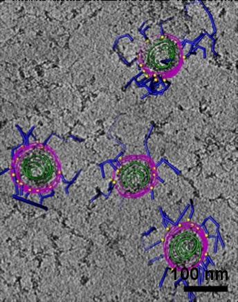
| Title | A Spiral Scaffold Underlies Cytoadherent Knobs in Plasmodium falciparum-Infected Erythrocytes | |||||||||
 Map data Map data | Cryo-tomogram of detergent-extracted cytoskeleton of P. falciparum-infected erythrocyte, schiztont stage, showing knob skeletons. Map has been NAD and high-pass filtered to remove background gradient. | |||||||||
 Sample Sample |
| |||||||||
 Keywords Keywords |  malaria / Plasmodium / knobs / cytoadherence / KAHRP / erythrocyte / skeleton / schizont / blood malaria / Plasmodium / knobs / cytoadherence / KAHRP / erythrocyte / skeleton / schizont / blood | |||||||||
| Biological species |  Plasmodium falciparum (malaria parasite P. falciparum) Plasmodium falciparum (malaria parasite P. falciparum) | |||||||||
| Method | electron tomography / cryo EM | |||||||||
 Authors Authors | Watermeyer JM / Hale VL / Hackett F / Clare DK / Cutts EE / Vakonakis I / Fleck RA / Blackman MJ / Saibil HR | |||||||||
 Citation Citation | Journal: Blood / Year: 2016 Title: A spiral scaffold underlies cytoadherent knobs in Plasmodium falciparum-infected erythrocytes. Authors: Jean M Watermeyer / Victoria L Hale / Fiona Hackett / Daniel K Clare / Erin E Cutts / Ioannis Vakonakis / Roland A Fleck / Michael J Blackman / Helen R Saibil /  Abstract: Much of the virulence of Plasmodium falciparum malaria is caused by cytoadherence of infected erythrocytes, which promotes parasite survival by preventing clearance in the spleen. Adherence is ...Much of the virulence of Plasmodium falciparum malaria is caused by cytoadherence of infected erythrocytes, which promotes parasite survival by preventing clearance in the spleen. Adherence is mediated by membrane protrusions known as knobs, whose formation depends on the parasite-derived, knob-associated histidine-rich protein (KAHRP). Knobs are required for cytoadherence under flow conditions, and they contain both KAHRP and the parasite-derived erythrocyte membrane protein PfEMP1. Using electron tomography, we have examined the 3-dimensional structure of knobs in detergent-insoluble skeletons of P falciparum 3D7 schizonts. We describe a highly organized knob skeleton composed of a spiral structure coated by an electron-dense layer underlying the knob membrane. This knob skeleton is connected by multiple links to the erythrocyte cytoskeleton. We used immuno-electron microscopy (EM) to locate KAHRP in these structures. The arrangement of membrane proteins in the knobs, visualized by high-resolution freeze-fracture scanning EM, is distinct from that in the surrounding erythrocyte membrane, with a structure at the apex that likely represents the adhesion site. Thus, erythrocyte knobs in P falciparum infection contain a highly organized skeleton structure underlying a specialized region of membrane. We propose that the spiral and dense coat organize the cytoadherence structures in the knob, and anchor them into the erythrocyte cytoskeleton. The high density of knobs and their extensive mechanical linkage suggest an explanation for the rigidification of the cytoskeleton in infected cells, and for the transmission to the cytoskeleton of shear forces experienced by adhering cells. | |||||||||
| History |
|
- Structure visualization
| Movie |
 Movie viewer Movie viewer |
|---|---|
| Structure viewer | EM map: SurfViewMolmilJmol/JSmol |
| Supplemental images |
- Downloads & links
-EMDB archive
| Map data | emd_3116.map.gz | 319.3 MB | EMDB map data format | |
|---|---|---|---|---|
| Header (meta data) | emd-3116-v30.xmlemd-3116.xml | 9.6 KB 9.6 KB | Display Display | EMDB header |
| Images | EMD-3116.tif | 348.5 KB | ||
| Archive directory |  http://ftp.pdbj.org/pub/emdb/structures/EMD-3116ftp://ftp.pdbj.org/pub/emdb/structures/EMD-3116 http://ftp.pdbj.org/pub/emdb/structures/EMD-3116ftp://ftp.pdbj.org/pub/emdb/structures/EMD-3116 | HTTPS FTP |
-Related structure data
-Links
| EMDB pages | EMDB (EBI/PDBe) / EMDataResource |
|---|
-Map
| File | Download / File: emd_3116.map.gz / Format: CCP4 / Size: 770 MB / Type: IMAGE STORED AS SIGNED INTEGER (2 BYTES) | ||||||||||||||||||||||||||||||||||||||||||||||||||||||||||||
|---|---|---|---|---|---|---|---|---|---|---|---|---|---|---|---|---|---|---|---|---|---|---|---|---|---|---|---|---|---|---|---|---|---|---|---|---|---|---|---|---|---|---|---|---|---|---|---|---|---|---|---|---|---|---|---|---|---|---|---|---|---|
| Annotation | Cryo-tomogram of detergent-extracted cytoskeleton of P. falciparum-infected erythrocyte, schiztont stage, showing knob skeletons. Map has been NAD and high-pass filtered to remove background gradient. | ||||||||||||||||||||||||||||||||||||||||||||||||||||||||||||
| Voxel size | X=Y=Z: 10.78 Å | ||||||||||||||||||||||||||||||||||||||||||||||||||||||||||||
| Density |
| ||||||||||||||||||||||||||||||||||||||||||||||||||||||||||||
| Symmetry | Space group: 1 | ||||||||||||||||||||||||||||||||||||||||||||||||||||||||||||
| Details | EMDB XML:
CCP4 map header:
| ||||||||||||||||||||||||||||||||||||||||||||||||||||||||||||
-Supplemental data
- Sample components
-Entire : Detergent-resistant skeleton of P. falciparum schizont
| Entire | Name: Detergent-resistant skeleton of P. falciparum schizont |
|---|---|
| Components |
|
-Supramolecule #1000: Detergent-resistant skeleton of P. falciparum schizont
| Supramolecule | Name: Detergent-resistant skeleton of P. falciparum schizont type: sample / ID: 1000 / Number unique components: 1 |
|---|
-Supramolecule #1: Cytoskeleton with knobs
| Supramolecule | Name: Cytoskeleton with knobs / type: organelle_or_cellular_component / ID: 1 Details: Mature schizonts were lysed and detergent-extracted on EM grids, then plunge-frozen. Recombinant expression: No / Database: NCBI |
|---|---|
| Source (natural) | Organism: Plasmodium falciparum (malaria parasite P. falciparum) Strain: 3D7 / synonym: malaria parasite / Cell: schizont / Location in cell: erythrocyte cytoskeleton |
-Experimental details
-Structure determination
| Method | cryo EM |
|---|---|
 Processing Processing | electron tomography |
| Aggregation state | cell |
-Sample preparation
| Buffer | pH: 7.4 |
|---|---|
| Grid | Details: 300 mesh copper grid with lacy carbon support and thin carbon overlay |
| Vitrification | Cryogen name: ETHANE / Instrument: HOMEMADE PLUNGER |
- Electron microscopy
| Microscope | FEI POLARA 300 |
|---|---|
| Electron beam | Acceleration voltage: 300 kV / Electron source: FIELD EMISSION GUN |
| Electron optics | Illumination mode: FLOOD BEAM / Imaging mode: BRIGHT FIELDBright-field microscopy / Cs: 2.3 mm / Nominal defocus max: 8.0 µm / Nominal defocus min: 8.0 µm |
| Specialist optics | Energy filter - Name: Gatan Quantum |
| Sample stage | Specimen holder model: SIDE ENTRY, EUCENTRIC / Tilt series - Axis1 - Min angle: -69 ° / Tilt series - Axis1 - Max angle: 53 ° / Tilt series - Axis1 - Angle increment: 2 ° |
| Details | counting mode |
| Date | Aug 1, 2014 |
| Image recording | Category: CCD / Film or detector model: GATAN K2 (4k x 4k) / Average electron dose: 53 e/Å2 Details: Images were the average of 6 subframes recorded by the direct electron detector |
| Experimental equipment |  Model: Tecnai Polara / Image courtesy: FEI Company |
-Image processing
| CTF correction | Details: each image |
|---|---|
| Final reconstruction | Software - Name: IMOD / Number images used: 62 |
