- EMDB-3088: Structural basis for DNA strand separation by a hexameric replica... -
+
Open data
ID or keywords:
Loading...
-
Basic information
Entry
Database: EMDB / ID: EMD-3088
Title
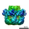
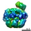
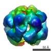
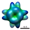
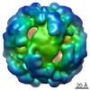
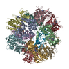
Structural basis for DNA strand separation by a hexameric replicative helicase
Map data
Non-symmetrised reconstruction of full length E1 helices from bovine papillomavirus
Sample
Sample: Full-length E1 helicase-DNA complex
Protein or peptide: Full-length hexameric E1 helicase
Keywords
papillomavirus / helicase / DNA replication fork / electron microscopy / structural analysis
Function / homology
Function and homology information
DNA helicase activity / DNA replication / DNA helicase / host cell nucleus / ATP hydrolysis activity / DNA binding / ATP binding Similarity search - Function
DNA helicase E1, C-terminal, Papillomavirus / DNA helicase E1, N-terminal, Papillomavirus / Replication protein E1, papillomavirus / DNA helicase E1, DNA-binding domain, papillomavirus / DNA helicase E1, DNA-binding domain superfamily, papillomavirus / Papillomavirus helicase / E1 Protein, N terminal domain / Papillomavirus E1, DNA-binding domain / Zinc finger, large T-antigen D1 domain superfamily / Helicase, superfamily 3, DNA virus ...DNA helicase E1, C-terminal, Papillomavirus / DNA helicase E1, N-terminal, Papillomavirus / Replication protein E1, papillomavirus / DNA helicase E1, DNA-binding domain, papillomavirus / DNA helicase E1, DNA-binding domain superfamily, papillomavirus / Papillomavirus helicase / E1 Protein, N terminal domain / Papillomavirus E1, DNA-binding domain / Zinc finger, large T-antigen D1 domain superfamily / Helicase, superfamily 3, DNA virus / Superfamily 3 helicase of DNA viruses domain profile. / P-loop containing nucleoside triphosphate hydrolase Similarity search - Domain/homology
Journal: Nucleic Acids Res / Year: 2015 Title: Structural basis for DNA strand separation by a hexameric replicative helicase. Authors: Yuriy Chaban / Jonathan A Stead / Ksenia Ryzhenkova / Fiona Whelan / Ekaterina P Lamber / Alfred Antson / Cyril M Sanders / Elena V Orlova / Abstract: Hexameric helicases are processive DNA unwinding machines but how they engage with a replication fork during unwinding is unknown. Using electron microscopy and single particle analysis we determined ...Hexameric helicases are processive DNA unwinding machines but how they engage with a replication fork during unwinding is unknown. Using electron microscopy and single particle analysis we determined structures of the intact hexameric helicase E1 from papillomavirus and two complexes of E1 bound to a DNA replication fork end-labelled with protein tags. By labelling a DNA replication fork with streptavidin (dsDNA end) and Fab (5' ssDNA) we located the positions of these labels on the helicase surface, showing that at least 10 bp of dsDNA enter the E1 helicase via a side tunnel. In the currently accepted 'steric exclusion' model for dsDNA unwinding, the active 3' ssDNA strand is pulled through a central tunnel of the helicase motor domain as the dsDNA strands are wedged apart outside the protein assembly. Our structural observations together with nuclease footprinting assays indicate otherwise: strand separation is taking place inside E1 in a chamber above the helicase domain and the 5' passive ssDNA strands exits the assembly through a separate tunnel opposite to the dsDNA entry point. Our data therefore suggest an alternative to the current general model for DNA unwinding by hexameric helicases.
History
Deposition
Jul 9, 2015
-
Header (metadata) release
Aug 5, 2015
-
Map release
Aug 19, 2015
-
Update
Oct 7, 2015
-
Current status
Oct 7, 2015
Processing site: PDBe / Status: Released
-
Structure visualization
Movie
Surface view with section colored by density value
pH: 8 Details: 10 mM Tris-Cl pH 8.0, 225 mM NaCl, 2 mM DTT, 0.1 mM PMSF, 0.1 mM EDTA
Staining
Type: NEGATIVE / Details: Sample was stained with 2% uranyl acetate
Grid
Details: Sample was applied on to carbon-coated copper grids (400 mesh, freshly glow-discharged in air)
Vitrification
Cryogen name: NONE / Instrument: OTHER
-
Electron microscopy
Microscope
FEI TECNAI F20
Temperature
Min: 291 K / Max: 296 K / Average: 293 K
Alignment procedure
Legacy - Astigmatism: Objective lens astigmatism was corrected at 100,000 times magnification
Date
Apr 15, 2012
Image recording
Category: CCD / Film or detector model: GATAN ULTRASCAN 4000 (4k x 4k) / Digitization - Sampling interval: 1.6 µm / Number real images: 45 / Average electron dose: 20 e/Å2 / Camera length: 1000 / Details: No subframe averaging was used. / Bits/pixel: 16
Electron beam
Acceleration voltage: 200 kV / Electron source: FIELD EMISSION GUN
Specimen holder: Negative stain holder / Specimen holder model: OTHER
Experimental equipment
Model: Tecnai F20 / Image courtesy: FEI Company
+
Image processing
Details
Particle picking was carried out automatically using BOXER software. Initial references were prepared using several manually selected protein complex images representing different views.
CTF correction
Details: frames
Final reconstruction
Applied symmetry - Point group: C1 (asymmetric) / Algorithm: OTHER / Resolution.type: BY AUTHOR / Resolution: 23.0 Å / Resolution method: OTHER / Software - Name: Imagic, CTFit, CTFFIND3 / Details: Final map was calculated from 500 classes / Number images used: 3553
In the structure databanks used in Yorodumi, some data are registered as the other names, "COVID-19 virus" and "2019-nCoV". Here are the details of the virus and the list of structure data.
Jan 31, 2019. EMDB accession codes are about to change! (news from PDBe EMDB page)
EMDB accession codes are about to change! (news from PDBe EMDB page)
The allocation of 4 digits for EMDB accession codes will soon come to an end. Whilst these codes will remain in use, new EMDB accession codes will include an additional digit and will expand incrementally as the available range of codes is exhausted. The current 4-digit format prefixed with “EMD-” (i.e. EMD-XXXX) will advance to a 5-digit format (i.e. EMD-XXXXX), and so on. It is currently estimated that the 4-digit codes will be depleted around Spring 2019, at which point the 5-digit format will come into force.
The EM Navigator/Yorodumi systems omit the EMD- prefix.
Related info.:Q: What is EMD? / ID/Accession-code notation in Yorodumi/EM Navigator
Yorodumi is a browser for structure data from EMDB, PDB, SASBDB, etc.
This page is also the successor to EM Navigator detail page, and also detail information page/front-end page for Omokage search.
The word "yorodu" (or yorozu) is an old Japanese word meaning "ten thousand". "mi" (miru) is to see.
Related info.:EMDB / PDB / SASBDB / Comparison of 3 databanks / Yorodumi Search / Aug 31, 2016. New EM Navigator & Yorodumi / Yorodumi Papers / Jmol/JSmol / Function and homology information / Changes in new EM Navigator and Yorodumi
 Movie
Movie Controller

 Yorodumi
 Open data
 Basic information
 Map data
 Sample
 Keywords
 Function and homology information
 Bovine papillomavirus
 Authors
 Citation
 Structure visualization
 Downloads & links
 emd_3088.png
 http://ftp.pdbj.org/pub/emdb/structures/EMD-3088


 Sample components
 Processing
 Electron microscopy
 FIELD EMISSION GUN


