Movie
Movie Controller

[English] 日本語
 Yorodumi
- EMDB-3075: Cyclophilin A Stabilizes HIV-1 Capsid through a Novel Non-canonic... -
+ Open data

- Basic information
| Entry | Database: EMDB / ID: EMD-3075 | |||||||||
|---|---|---|---|---|---|---|---|---|---|---|
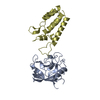
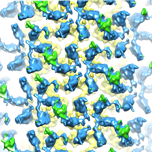
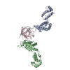
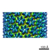
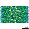
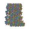
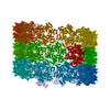
| Title | Cyclophilin A Stabilizes HIV-1 Capsid through a Novel Non-canonical Binding Site | |||||||||
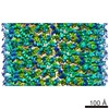
 Map data Map data | Reconstruction of HIV-1 capsid with Cypa | |||||||||
 Sample Sample |
| |||||||||
| Biological species |   Human immunodeficiency virus 1 Human immunodeficiency virus 1 | |||||||||
| Method | helical reconstruction / cryo EM / Resolution: 8.0 Å | |||||||||
 Authors Authors | Liu C / Perilla JR / Ning J / Lu M / Hou G / Ramalhu R / Bedwell GJ / Ahn J / Shi J / Gronenborn AM ...Liu C / Perilla JR / Ning J / Lu M / Hou G / Ramalhu R / Bedwell GJ / Ahn J / Shi J / Gronenborn AM / Prevelige Jr PE / Rousso I / Aiken C / Polenova T / Schulten K / Zhang P | |||||||||
 Citation Citation | Journal: Nat Commun / Year: 2016 Title: Cyclophilin A stabilizes the HIV-1 capsid through a novel non-canonical binding site. Authors: Chuang Liu / Juan R Perilla / Jiying Ning / Manman Lu / Guangjin Hou / Ruben Ramalho / Benjamin A Himes / Gongpu Zhao / Gregory J Bedwell / In-Ja Byeon / Jinwoo Ahn / Angela M Gronenborn / ...Authors: Chuang Liu / Juan R Perilla / Jiying Ning / Manman Lu / Guangjin Hou / Ruben Ramalho / Benjamin A Himes / Gongpu Zhao / Gregory J Bedwell / In-Ja Byeon / Jinwoo Ahn / Angela M Gronenborn / Peter E Prevelige / Itay Rousso / Christopher Aiken / Tatyana Polenova / Klaus Schulten / Peijun Zhang /   Abstract: The host cell factor cyclophilin A (CypA) interacts directly with the HIV-1 capsid and regulates viral infectivity. Although the crystal structure of CypA in complex with the N-terminal domain of the ...The host cell factor cyclophilin A (CypA) interacts directly with the HIV-1 capsid and regulates viral infectivity. Although the crystal structure of CypA in complex with the N-terminal domain of the HIV-1 capsid protein (CA) has been known for nearly two decades, how CypA interacts with the viral capsid and modulates HIV-1 infectivity remains unclear. We determined the cryoEM structure of CypA in complex with the assembled HIV-1 capsid at 8-Å resolution. The structure exhibits a distinct CypA-binding pattern in which CypA selectively bridges the two CA hexamers along the direction of highest curvature. EM-guided all-atom molecular dynamics simulations and solid-state NMR further reveal that the CypA-binding pattern is achieved by single-CypA molecules simultaneously interacting with two CA subunits, in different hexamers, through a previously uncharacterized non-canonical interface. These results provide new insights into how CypA stabilizes the HIV-1 capsid and is recruited to facilitate HIV-1 infection. | |||||||||
| History |
|
- Structure visualization
| Movie |
 Movie viewer Movie viewer |
|---|---|
| Structure viewer | EM map: SurfViewMolmilJmol/JSmol |
| Supplemental images |
- Downloads & links
-EMDB archive
| Map data | emd_3075.map.gz | 91.4 MB | EMDB map data format | |
|---|---|---|---|---|
| Header (meta data) | emd-3075-v30.xmlemd-3075.xml | 11.6 KB 11.6 KB | Display Display | EMDB header |
| Images |  EMDB_3075.png EMDB_3075.png | 389.3 KB | ||
| Archive directory |  http://ftp.pdbj.org/pub/emdb/structures/EMD-3075ftp://ftp.pdbj.org/pub/emdb/structures/EMD-3075 http://ftp.pdbj.org/pub/emdb/structures/EMD-3075ftp://ftp.pdbj.org/pub/emdb/structures/EMD-3075 | HTTPS FTP |
-Validation report
| Summary document | emd_3075_validation.pdf.gz | 241.4 KB | Display | EMDB validaton report |
|---|---|---|---|---|
| Full document | emd_3075_full_validation.pdf.gz | 240.5 KB | Display | |
| Data in XML | emd_3075_validation.xml.gz | 5 KB | Display | |
| Arichive directory | https://ftp.pdbj.org/pub/emdb/validation_reports/EMD-3075ftp://ftp.pdbj.org/pub/emdb/validation_reports/EMD-3075 | HTTPS FTP |
-Related structure data
-Links
| EMDB pages | EMDB (EBI/PDBe) / EMDataResource |
|---|
-Map
| File | Download / File: emd_3075.map.gz / Format: CCP4 / Size: 184.1 MB / Type: IMAGE STORED AS FLOATING POINT NUMBER (4 BYTES) | ||||||||||||||||||||||||||||||||||||||||||||||||||||||||||||||||||||
|---|---|---|---|---|---|---|---|---|---|---|---|---|---|---|---|---|---|---|---|---|---|---|---|---|---|---|---|---|---|---|---|---|---|---|---|---|---|---|---|---|---|---|---|---|---|---|---|---|---|---|---|---|---|---|---|---|---|---|---|---|---|---|---|---|---|---|---|---|---|
| Annotation | Reconstruction of HIV-1 capsid with Cypa | ||||||||||||||||||||||||||||||||||||||||||||||||||||||||||||||||||||
| Voxel size | X=Y=Z: 1.06 Å | ||||||||||||||||||||||||||||||||||||||||||||||||||||||||||||||||||||
| Density |
| ||||||||||||||||||||||||||||||||||||||||||||||||||||||||||||||||||||
| Symmetry | Space group: 1 | ||||||||||||||||||||||||||||||||||||||||||||||||||||||||||||||||||||
| Details | EMDB XML:
CCP4 map header:
| ||||||||||||||||||||||||||||||||||||||||||||||||||||||||||||||||||||
-Supplemental data
- Sample components
-Entire : Helical assembly of HIV-1 capsid protein and host cell factor Cyc...
| Entire | Name: Helical assembly of HIV-1 capsid protein and host cell factor Cyclophilin A |
|---|---|
| Components |
|
-Supramolecule #1000: Helical assembly of HIV-1 capsid protein and host cell factor Cyc...
| Supramolecule | Name: Helical assembly of HIV-1 capsid protein and host cell factor Cyclophilin A type: sample / ID: 1000 / Oligomeric state: hexamer / Number unique components: 1 |
|---|---|
| Molecular weight | Experimental: 42 KDa / Theoretical: 42 KDa |
-Macromolecule #1: Human immunodeficiency virus 1
| Macromolecule | Name: Human immunodeficiency virus 1 / type: protein_or_peptide / ID: 1 / Name.synonym: HIV-1 capsid protein with CypA / Oligomeric state: Hexamer / Recombinant expression: Yes |
|---|---|
| Source (natural) | Organism: Human immunodeficiency virus 1 |
| Molecular weight | Experimental: 42 KDa / Theoretical: 42 KDa |
| Recombinant expression | Organism:  |
-Experimental details
-Structure determination
| Method | cryo EM |
|---|---|
 Processing Processing | helical reconstruction |
| Aggregation state | helical array |
-Sample preparation
| Concentration | 2 mg/mL |
|---|---|
| Buffer | pH: 8 / Details: 1M NaCl,50mM Tris-Hcl |
| Grid | Details: Glow discharged perforated Quantifoil R2/1 grid |
| Vitrification | Cryogen name: ETHANE / Chamber humidity: 100 % / Chamber temperature: 120 K / Instrument: HOMEMADE PLUNGER Method: With 2.5 uL sample on carbon side, add 3 uL dilution buffer (100 mM NaCl, 50 mM Tris, pH 8.0) to back side. Blot 3-5 seconds from back side. |
- Electron microscopy
| Microscope | FEI TECNAI F20 |
|---|---|
| Date | Jun 20, 2013 |
| Image recording | Category: FILM / Film or detector model: KODAK SO-163 FILM / Digitization - Scanner: NIKON SUPER COOLSCAN 9000 / Digitization - Sampling interval: 6.349 µm / Number real images: 19 / Average electron dose: 15 e/Å2 / Bits/pixel: 32 |
| Electron beam | Acceleration voltage: 200 kV / Electron source:  FIELD EMISSION GUN FIELD EMISSION GUN |
| Electron optics | Illumination mode: FLOOD BEAM / Imaging mode: BRIGHT FIELD / Cs: 2.0 mm / Nominal defocus max: 3.5 µm / Nominal defocus min: 1.0 µm / Nominal magnification: 59000 |
| Sample stage | Specimen holder model: GATAN LIQUID NITROGEN |
| Experimental equipment |  Model: Tecnai F20 / Image courtesy: FEI Company |
-Image processing
| Details | The particles were aligned using IHRSR |
|---|---|
| Final reconstruction | Applied symmetry - Helical parameters - Δz: 7.393 Å Applied symmetry - Helical parameters - Δ&Phi: 138.133 ° Applied symmetry - Helical parameters - Axial symmetry: C1 (asymmetric) Resolution.type: BY AUTHOR / Resolution: 8.0 Å / Resolution method: OTHER / Software - Name: SPIDER, IHRSR |
| CTF correction | Details: Each particle |
-Atomic model buiding 1
| Initial model | PDB ID: |
|---|---|
| Software | Name: Chimera |
| Refinement | Space: REAL / Protocol: RIGID BODY FIT |
-Atomic model buiding 2
| Initial model | PDB ID: |
|---|---|
| Software | Name: Chimera |
| Refinement | Space: REAL / Protocol: RIGID BODY FIT |