 Movie
Movie Controller

[English] 日本語
 Yorodumi
- EMDB-2823: Structure determination of feline calicivirus virus-like particle... -
+ Open data

- Basic information
| Entry | Database: EMDB / ID: EMD-2823 | |||||||||
|---|---|---|---|---|---|---|---|---|---|---|
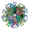
| Title | Structure determination of feline calicivirus virus-like particles in the context of a pseudo-octahedral arrangement | |||||||||
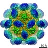
 Map data Map data | Reconstruction of feline calicivirus VLP T=1. | |||||||||
 Sample Sample |
| |||||||||
 Keywords Keywords |  virus-like particle / VLP / VP1 / calicivirus / FCV / icosahedral symmetry / octahedral symmetry / non-crystallographic symmetry / feline calici virus / vaccine / subviral particle virus-like particle / VLP / VP1 / calicivirus / FCV / icosahedral symmetry / octahedral symmetry / non-crystallographic symmetry / feline calici virus / vaccine / subviral particle | |||||||||
| Function / homology | Calicivirus coat protein / Calicivirus coat protein / T=3 icosahedral viral capsid / Picornavirus/Calicivirus coat protein / Viral coat protein subunit / Capsid protein Function and homology information Function and homology information | |||||||||
| Biological species |  Canine calicivirus Canine calicivirus | |||||||||
| Method | single particle reconstruction / cryo EM / negative staining / Resolution: 14.0 Å | |||||||||
 Authors Authors | Burmeister WP / Buisson M / Estrozi LF / Schoehn G / Billet O / Hannas Z / Sigoillot-Claude C / Poulet H | |||||||||
 Citation Citation | Journal: PLoS One / Year: 2015 Title: Structure determination of feline calicivirus virus-like particles in the context of a pseudo-octahedral arrangement. Authors: Wim P Burmeister / Marlyse Buisson / Leandro F Estrozi / Guy Schoehn / Olivier Billet / Zahia Hannas / Cécile Sigoillot / Hervé Poulet /  Abstract: The vesivirus feline calicivirus (FCV) is a positive strand RNA virus encapsidated by an icosahedral T=3 shell formed by the viral VP1 protein. Upon its expression in the insect cell - baculovirus ...The vesivirus feline calicivirus (FCV) is a positive strand RNA virus encapsidated by an icosahedral T=3 shell formed by the viral VP1 protein. Upon its expression in the insect cell - baculovirus system in the context of vaccine development, two types of virus-like particles (VLPs) were formed, a majority built of 60 subunits (T=1) and a minority probably built of 180 subunits (T=3). The structure of the small particles was determined by x-ray crystallography at 0.8 nm resolution helped by cryo-electron microscopy in order to understand their formation. Cubic crystals belonged to space group P213. Their self-rotation function showed the presence of an octahedral pseudo-symmetry similar to the one described previously by Agerbandje and co-workers for human parvovirus VLPs. The crystal structure could be solved starting from the published VP1 structure in the context of the T=3 viral capsid. In contrast to viral capsids, where the capsomers are interlocked by the exchange of the N-terminal arm (NTA) domain, this domain is disordered in the T=1 capsid of the VLPs. Furthermore it is prone to proteolytic cleavage. The relative orientation of P (protrusion) and S (shell) domains is alerted so as to fit VP1 to the smaller T=1 particle whereas the intermolecular contacts around 2-fold, 3-fold and 5-fold axes are conserved. By consequence the surface of the VLP is very similar compared to the viral capsid and suggests a similar antigenicity. The knowledge of the structure of the VLPs will help to improve their stability, in respect to a use for vaccination. | |||||||||
| History |
|
- Structure visualization
| Movie |
Movie viewer |
|---|---|
| Structure viewer | EM map: SurfViewMolmilJmol/JSmol |
- Downloads & links
-EMDB archive
| Map data | emd_2823.map.gz | 12 MB | EMDB map data format | |
|---|---|---|---|---|
| Header (meta data) | emd-2823-v30.xmlemd-2823.xml | 9.2 KB 9.2 KB | Display Display | EMDB header |
| Images |  EMD-2823.png EMD-2823.png | 173.2 KB | ||
| Archive directory |  http://ftp.pdbj.org/pub/emdb/structures/EMD-2823ftp://ftp.pdbj.org/pub/emdb/structures/EMD-2823 http://ftp.pdbj.org/pub/emdb/structures/EMD-2823ftp://ftp.pdbj.org/pub/emdb/structures/EMD-2823 | HTTPS FTP |
-Related structure data


-Links
| EMDB pages | EMDB (EBI/PDBe) / EMDataResource |
|---|---|
| Related items in Molecule of the Month |


-Map
| File | Download / File: emd_2823.map.gz / Format: CCP4 / Size: 12.6 MB / Type: IMAGE STORED AS FLOATING POINT NUMBER (4 BYTES) | ||||||||||||||||||||||||||||||||||||||||||||||||||||||||||||
|---|---|---|---|---|---|---|---|---|---|---|---|---|---|---|---|---|---|---|---|---|---|---|---|---|---|---|---|---|---|---|---|---|---|---|---|---|---|---|---|---|---|---|---|---|---|---|---|---|---|---|---|---|---|---|---|---|---|---|---|---|---|
| Annotation | Reconstruction of feline calicivirus VLP T=1. | ||||||||||||||||||||||||||||||||||||||||||||||||||||||||||||
| Voxel size | X=Y=Z: 2.33333 Å | ||||||||||||||||||||||||||||||||||||||||||||||||||||||||||||
| Density |
| ||||||||||||||||||||||||||||||||||||||||||||||||||||||||||||
| Symmetry | Space group: 1 | ||||||||||||||||||||||||||||||||||||||||||||||||||||||||||||
| Details | EMDB XML:
CCP4 map header:
| ||||||||||||||||||||||||||||||||||||||||||||||||||||||||||||
-Supplemental data
- Sample components
-Entire : feline calicivirus virus-like particles
| Entire | Name: feline calicivirus virus-like particles |
|---|---|
| Components |
|
-Supramolecule #1000: feline calicivirus virus-like particles
| Supramolecule | Name: feline calicivirus virus-like particles / type: sample / ID: 1000 / Oligomeric state: icosahedral / Number unique components: 1 |
|---|---|
| Molecular weight | Theoretical: 3.552 MDa |
-Supramolecule #1: Canine calicivirus
| Supramolecule | Name: Canine calicivirus / type: virus / ID: 1 / NCBI-ID: 74724 / Sci species name: Canine calicivirus / Virus type: VIRUS-LIKE PARTICLE / Virus isolate: OTHER / Virus enveloped: No / Virus empty: Yes |
|---|---|
| Host (natural) | Organism:  Felis catus (domestic cat) / synonym: VERTEBRATES Felis catus (domestic cat) / synonym: VERTEBRATES |
| Host system | Organism:   Spodoptera frugiperda (fall armyworm) / Recombinant cell: Sf9 / Recombinant plasmid: Merial100869 Spodoptera frugiperda (fall armyworm) / Recombinant cell: Sf9 / Recombinant plasmid: Merial100869 |
| Molecular weight | Theoretical: 3.5 MDa |
| Virus shell | Shell ID: 1 / Diameter: 280 Å / T number (triangulation number): 1 |
-Experimental details
-Structure determination
| Method | negative staining, cryo EM |
|---|---|
 Processing Processing | single particle reconstruction |
| Aggregation state | particle |
-Sample preparation
| Buffer | pH: 7 / Details: 20 mM MES pH 6, 200 mM NaCl |
|---|---|
| Staining | Type: NEGATIVE / Details: no stain (cryo-EM) |
| Grid | Details: Quantifoil R2/1 holey grid |
| Vitrification | Cryogen name: ETHANE / Instrument: FEI VITROBOT MARK IV |
- Electron microscopy
| Microscope | FEI POLARA 300 |
|---|---|
| Electron beam | Acceleration voltage: 300 kV / Electron source: FIELD EMISSION GUN |
| Electron optics | Illumination mode: FLOOD BEAM / Imaging mode: BRIGHT FIELDBright-field microscopy / Cs: 2.0 mm / Nominal defocus max: 3.0 µm / Nominal defocus min: 1.0 µm / Nominal magnification: 39000 |
| Sample stage | Specimen holder: Top-entry polara / Specimen holder model: OTHER |
| Date | Sep 1, 2010 |
| Image recording | Category: FILM / Film or detector model: KODAK SO-163 FILM / Digitization - Scanner: ZEISS SCAI / Digitization - Sampling interval: 7 µm / Number real images: 20 / Average electron dose: 20 e/Å2 / Bits/pixel: 8 |
| Experimental equipment |  Model: Tecnai Polara / Image courtesy: FEI Company |
-Image processing
| CTF correction | Details: Phase-flipping |
|---|---|
| Final reconstruction | Applied symmetry - Point group: I (icosahedral) / Algorithm: OTHER / Resolution.type: BY AUTHOR / Resolution: 14.0 Å / Resolution method: OTHER / Software - Name: RIco, Bsoft, ctffind3, FPM / Number images used: 11158 |
| Details | Particles semi-automatically picked by the boxer program (EMAN) |
