 Movie
Movie Controller

[English] 日本語
 Yorodumi
- EMDB-2790: The molecular structure of the left-handed supra- molecular helix... -
+ Open data

- Basic information
| Entry | Database: EMDB / ID: EMD-2790 | |||||||||
|---|---|---|---|---|---|---|---|---|---|---|
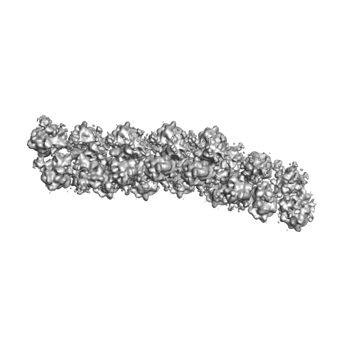
| Title | The molecular structure of the left-handed supra- molecular helix of eukaryotic polyribosomes | |||||||||
 Map data Map data | 3D poly-ribosome structure from Wheat Germ in vitro cell free system | |||||||||
 Sample Sample |
| |||||||||
 Keywords Keywords | cryo-ET /  polyribosome / sub-tomogram averaging polyribosome / sub-tomogram averaging | |||||||||
| Function / homology |  Function and homology information Function and homology informationtranslational elongation / protein kinase activator activity / ribonucleoprotein complex binding / maturation of LSU-rRNA from tricistronic rRNA transcript (SSU-rRNA, 5.8S rRNA, LSU-rRNA) / maturation of LSU-rRNA / cytosolic ribosome / cytosolic small ribosomal subunit / large ribosomal subunit rRNA binding / small ribosomal subunit / cytosolic large ribosomal subunit ...translational elongation / protein kinase activator activity / ribonucleoprotein complex binding / maturation of LSU-rRNA from tricistronic rRNA transcript (SSU-rRNA, 5.8S rRNA, LSU-rRNA) / maturation of LSU-rRNA / cytosolic ribosome / cytosolic small ribosomal subunit / large ribosomal subunit rRNA binding / small ribosomal subunit / cytosolic large ribosomal subunit / cytoplasmic translation / negative regulation of translation / ribosome / structural constituent of ribosome / translation / ribonucleoprotein complex / mRNA binding / signal transduction / RNA binding / zinc ion binding / cytoplasmSimilarity search - Function | |||||||||
| Biological species |  Triticum aestivum (bread wheat) Triticum aestivum (bread wheat) | |||||||||
| Method | subtomogram averaging / cryo EM / Resolution: 34.0 Å | |||||||||
 Authors Authors | Myasnikov AG / Afonina ZHA / Menetret J-F / Shirokov VA / Spirin AS / Klaholz BP | |||||||||
 Citation Citation | Journal: Nat Commun / Year: 2014 Title: The molecular structure of the left-handed supra-molecular helix of eukaryotic polyribosomes. Authors: Alexander G Myasnikov / Zhanna A Afonina / Jean-François Ménétret / Vladimir A Shirokov / Alexander S Spirin / Bruno P Klaholz /   Abstract: During protein synthesis, several ribosomes bind to a single messenger RNA (mRNA) forming large macromolecular assemblies called polyribosomes. Here we report the detailed molecular structure of a ...During protein synthesis, several ribosomes bind to a single messenger RNA (mRNA) forming large macromolecular assemblies called polyribosomes. Here we report the detailed molecular structure of a 100 MDa eukaryotic poly-ribosome complex derived from cryo electron tomography, sub-tomogram averaging and pseudo-atomic modelling by crystal structure fitting. The structure allowed the visualization of the three functional parts of the polysome assembly, the central core region that forms a rather compact left-handed supra-molecular helix, and the more open regions that harbour the initiation and termination sites at either ends. The helical region forms a continuous mRNA channel where the mRNA strand bridges neighbouring exit and entry sites of the ribosomes and prevents mRNA looping between ribosomes. This structure provides unprecedented insights into protein- and RNA-mediated inter-ribosome contacts that involve conserved sites through 40S subunits and long protruding RNA expansion segments, suggesting a role in stabilizing the overall polyribosomal assembly. | |||||||||
| History |
|
- Structure visualization
| Movie |
Movie viewer |
|---|---|
| Structure viewer | EM map: SurfViewMolmilJmol/JSmol |
| Supplemental images |
- Downloads & links
-EMDB archive
| Map data | emd_2790.map.gz | 32.5 MB | EMDB map data format | |
|---|---|---|---|---|
| Header (meta data) | emd-2790-v30.xmlemd-2790.xml | 12.9 KB 12.9 KB | Display Display | EMDB header |
| Images |  EMD-2790.png EMD-2790.png | 70.4 KB | ||
| Archive directory |  http://ftp.pdbj.org/pub/emdb/structures/EMD-2790ftp://ftp.pdbj.org/pub/emdb/structures/EMD-2790 http://ftp.pdbj.org/pub/emdb/structures/EMD-2790ftp://ftp.pdbj.org/pub/emdb/structures/EMD-2790 | HTTPS FTP |
-Related structure data
| Related structure data | 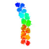 4v3pMC M: atomic model generated by this map C: citing same article ( |
|---|---|
| Similar structure data |

-Links
| EMDB pages | EMDB (EBI/PDBe) / EMDataResource |
|---|---|
| Related items in Molecule of the Month |


-Map
| File | Download / File: emd_2790.map.gz / Format: CCP4 / Size: 48.8 MB / Type: IMAGE STORED AS FLOATING POINT NUMBER (4 BYTES) | ||||||||||||||||||||||||||||||||||||||||||||||||||||||||||||||||||||
|---|---|---|---|---|---|---|---|---|---|---|---|---|---|---|---|---|---|---|---|---|---|---|---|---|---|---|---|---|---|---|---|---|---|---|---|---|---|---|---|---|---|---|---|---|---|---|---|---|---|---|---|---|---|---|---|---|---|---|---|---|---|---|---|---|---|---|---|---|---|
| Annotation | 3D poly-ribosome structure from Wheat Germ in vitro cell free system | ||||||||||||||||||||||||||||||||||||||||||||||||||||||||||||||||||||
| Voxel size | X=Y=Z: 6.8 Å | ||||||||||||||||||||||||||||||||||||||||||||||||||||||||||||||||||||
| Density |
| ||||||||||||||||||||||||||||||||||||||||||||||||||||||||||||||||||||
| Symmetry | Space group: 1 | ||||||||||||||||||||||||||||||||||||||||||||||||||||||||||||||||||||
| Details | EMDB XML:
CCP4 map header:
| ||||||||||||||||||||||||||||||||||||||||||||||||||||||||||||||||||||
-Supplemental data
- Sample components
-Entire : poly-ribosome from in vitro wheat germ system
| Entire | Name: poly-ribosome from in vitro wheat germ system |
|---|---|
| Components |
|
-Supramolecule #1000: poly-ribosome from in vitro wheat germ system
| Supramolecule | Name: poly-ribosome from in vitro wheat germ system / type: sample / ID: 1000 / Oligomeric state: 23-meric / Number unique components: 1 |
|---|---|
| Molecular weight | Theoretical: 100 MDa |
-Supramolecule #1: Wheat germ poly-ribosome
| Supramolecule | Name: Wheat germ poly-ribosome / type: complex / ID: 1 / Recombinant expression: No / Database: NCBI / Ribosome-details: ribosome-eukaryote: ALL |
|---|---|
| Source (natural) | Organism: Triticum aestivum (bread wheat) / synonym: Wheat Germ / Tissue: Germ |
| Molecular weight | Theoretical: 100 MDa |
-Experimental details
-Structure determination
| Method | cryo EM |
|---|---|
 Processing Processing | subtomogram averaging |
| Aggregation state | helical array |
-Sample preparation
| Concentration | 0.5 mg/mL |
|---|---|
| Buffer | pH: 7.6 Details: 25mM HEPES-KOH, 3mM Mg(OAc)2, 85mM KOAc, 1.6mM DTT, 0.25mM spermidine |
| Grid | Details: 3ul of sample applied on 300 mesh holy carbon Quantifoil 2/2 grid. Blotting was done in Vitrobot Mark IV |
| Vitrification | Cryogen name: ETHANE / Chamber humidity: 95 % / Chamber temperature: 120 K / Instrument: FEI VITROBOT MARK IV Method: 3ul of sample applied on 300 mesh holy carbon Quantifoil 2/2 grid. Blotting was done in Vitrobot Mark IV, blot time 0.5 sec, blot force 5 |
- Electron microscopy
| Microscope | FEI TECNAI F30 |
|---|---|
| Electron beam | Acceleration voltage: 150 kV / Electron source: FIELD EMISSION GUN |
| Electron optics | Calibrated magnification: 41176 / Illumination mode: FLOOD BEAM / Imaging mode: BRIGHT FIELDBright-field microscopy / Cs: 2 mm / Nominal defocus max: 4.0 µm / Nominal defocus min: 2.0 µm / Nominal magnification: 39000 |
| Sample stage | Specimen holder model: GATAN HELIUM / Tilt series - Axis1 - Min angle: -70 ° / Tilt series - Axis1 - Max angle: 70 ° |
| Temperature | Min: 80 K / Max: 100 K / Average: 90 K |
| Alignment procedure | Legacy - Astigmatism: Objective lens astigmatism was corrected at 120,000 times magnification Legacy - Electron beam tilt params: 0 |
| Date | Jan 1, 2013 |
| Image recording | Category: CCD / Film or detector model: FEI FALCON I (4k x 4k) / Average electron dose: 30 e/Å2 / Bits/pixel: 16 |
| Experimental equipment |  Model: Tecnai F30 / Image courtesy: FEI Company |
-Image processing
| Final 3D classification | Number classes: 1 |
|---|---|
| Final reconstruction | Applied symmetry - Helical parameters - Δz: 83 Å Applied symmetry - Helical parameters - Δ&Phi: 90 ° Applied symmetry - Helical parameters - Axial symmetry: C4 (4 fold cyclic )Algorithm: OTHER / Resolution.type: BY AUTHOR / Resolution: 34.0 Å / Resolution method: OTHER / Software - Name: Imod, Xmipp, Imagic / Number subtomograms used: 106 |
| Details | The subtomograms were selected in imod manually. The averaging was done in xmipp program by using ml_tomo subroutine. |
-Atomic model buiding 1
| Initial model | PDB ID:  3iz6 |
|---|---|
| Software | Name: Chimera |
| Details | 40S and 60S was fitted separately |
| Refinement | Space: REAL / Protocol: RIGID BODY FIT |
| Output model | PDB-4v3p: |
-Atomic model buiding 2
| Initial model | PDB ID: 3iz7 |
|---|---|
| Software | Name: Chimera |
| Details | 40S and 60S was fitted separately |
| Refinement | Space: REAL / Protocol: RIGID BODY FIT |
| Output model | PDB-4v3p: |
-Atomic model buiding 3
| Initial model | PDB ID: 3izr |
|---|---|
| Software | Name: Chimera |
| Details | 40S and 60S was fitted separately |
| Refinement | Space: REAL / Protocol: RIGID BODY FIT |
| Output model | PDB-4v3p: |
