Journal: J Biol Chem / Year: 2014 Title: Systematic comparison of molecular conformations of H+,K+-ATPase reveals an important contribution of the A-M2 linker for the luminal gating. Authors: Kazuhiro Abe / Kazutoshi Tani / Yoshinori Fujiyoshi / Abstract: Gastric H(+),K(+)-ATPase, an ATP-driven proton pump responsible for gastric acidification, is a molecular target for anti-ulcer drugs. Here we show its cryo-electron microscopy (EM) structure in an ...Gastric H(+),K(+)-ATPase, an ATP-driven proton pump responsible for gastric acidification, is a molecular target for anti-ulcer drugs. Here we show its cryo-electron microscopy (EM) structure in an E2P analog state, bound to magnesium fluoride (MgF), and its K(+)-competitive antagonist SCH28080, determined at 7 Å resolution by electron crystallography of two-dimensional crystals. Systematic comparison with other E2P-related cryo-EM structures revealed that the molecular conformation in the (SCH)E2·MgF state is remarkably distinguishable. Although the azimuthal position of the A domain of the (SCH)E2·MgF state is similar to that in the E2·AlF (aluminum fluoride) state, in which the transmembrane luminal gate is closed, the arrangement of transmembrane helices in the (SCH)E2·MgF state shows a luminal-open conformation imposed on by bound SCH28080 at its luminal cavity, based on observations of the structure in the SCH28080-bound E2·BeF (beryllium fluoride) state. The molecular conformation of the (SCH)E2·MgF state thus represents a mixed overall structure in which its cytoplasmic and luminal half appear to be independently modulated by a phosphate analog and an antagonist bound to the respective parts of the enzyme. Comparison of the molecular conformations revealed that the linker region connecting the A domain and the transmembrane helix 2 (A-M2 linker) mediates the regulation of luminal gating. The mechanistic rationale underlying luminal gating observed in H(+),K(+)-ATPase is consistent with that observed in sarcoplasmic reticulum Ca(2+)-ATPase and other P-type ATPases and is most likely conserved for the P-type ATPase family in general.
History
Deposition
Aug 18, 2014
-
Header (metadata) release
Sep 10, 2014
-
Map release
Sep 17, 2014
-
Update
Nov 12, 2014
-
Current status
Nov 12, 2014
Processing site: PDBe / Status: Released
-
Structure visualization
Movie
Surface view with section colored by density value
Name: H+,K+-ATPase / type: protein_or_peptide / ID: 1 / Number of copies: 2 / Oligomeric state: One alpha and one beta chain of HK-ATPase / Recombinant expression: No / Database: NCBI
Source (natural)
Organism: Sus scrofa (pig) / synonym: Pig / Tissue: gastric / Location in cell: Plasma membrane
Molecular weight
Theoretical: 110 KDa
Sequence
UniProtKB: Potassium-transporting ATPase alpha chain 1 / GO: ATP biosynthetic process / InterPro: P-type ATPase, A domain superfamily
-
Experimental details
-
Structure determination
Method
cryo EM
Processing
electron crystallography
Aggregation state
2D array
-
Sample preparation
Concentration
8 mg/mL
Buffer
pH: 4.8 Details: 20 mM propionate, 1 mM MgCl2, 1 mM AlCl3, 4 mM NaF, 1 mM ADP, 3 mM DTT and 10 M SCH28080 at pH 4.8 with Tris.
Grid
Details: molybdenum grid with thin carbon support
Vitrification
Cryogen name: NITROGEN / Instrument: LEICA KF80 Details: Vitrification carried out in cold room at 4 degrees Celsius
Details
Crystals grown in dialysis
Crystal formation
Details: Crystals grown in dialysis
-
Electron microscopy
Microscope
JEOL KYOTO-3000SFF
Electron beam
Acceleration voltage: 300 kV / Electron source: FIELD EMISSION GUN
Specimen holder: Helium cooled / Specimen holder model: JEOL / Tilt angle min: -63.6 / Tilt angle max: 63.6 / Tilt series - Axis1 - Min angle: -63.6 ° / Tilt series - Axis1 - Max angle: 63.6 °
Temperature
Min: 4 K / Average: 4 K
Date
Oct 29, 2013
Image recording
Category: FILM / Film or detector model: KODAK SO-163 FILM / Digitization - Scanner: ZEISS SCAI / Digitization - Sampling interval: 7 µm / Number real images: 267 / Average electron dose: 20 e/Å2 / Bits/pixel: 14
-
Image processing
Crystal parameters
Unit cell - A: 140.9 Å / Unit cell - B: 111.3 Å / Unit cell - C: 320.0 Å / Unit cell - γ: 90.0 ° / Unit cell - α: 90.0 ° / Unit cell - β: 90.0 ° / Plane group: P 2 21 21
CTF correction
Details: Each micrographs
Final reconstruction
Resolution.type: BY AUTHOR / Resolution: 8.0 Å / Resolution method: DIFFRACTION PATTERN/LAYERLINES / Software - Name: MRC
Details
Images were processed using MRC suite.
+
About Yorodumi
-
News
-
Feb 9, 2022. New format data for meta-information of EMDB entries
New format data for meta-information of EMDB entries
Version 3 of the EMDB header file is now the official format.
The previous official version 1.9 will be removed from the archive.
In the structure databanks used in Yorodumi, some data are registered as the other names, "COVID-19 virus" and "2019-nCoV". Here are the details of the virus and the list of structure data.
Jan 31, 2019. EMDB accession codes are about to change! (news from PDBe EMDB page)
EMDB accession codes are about to change! (news from PDBe EMDB page)
The allocation of 4 digits for EMDB accession codes will soon come to an end. Whilst these codes will remain in use, new EMDB accession codes will include an additional digit and will expand incrementally as the available range of codes is exhausted. The current 4-digit format prefixed with “EMD-” (i.e. EMD-XXXX) will advance to a 5-digit format (i.e. EMD-XXXXX), and so on. It is currently estimated that the 4-digit codes will be depleted around Spring 2019, at which point the 5-digit format will come into force.
The EM Navigator/Yorodumi systems omit the EMD- prefix.
Related info.:Q: What is EMD? / ID/Accession-code notation in Yorodumi/EM Navigator
Yorodumi is a browser for structure data from EMDB, PDB, SASBDB, etc.
This page is also the successor to EM Navigator detail page, and also detail information page/front-end page for Omokage search.
The word "yorodu" (or yorozu) is an old Japanese word meaning "ten thousand". "mi" (miru) is to see.
Related info.:EMDB / PDB / SASBDB / Comparison of 3 databanks / Yorodumi Search / Aug 31, 2016. New EM Navigator & Yorodumi / Yorodumi Papers / Jmol/JSmol / Function and homology information / Changes in new EM Navigator and Yorodumi
 Movie
Movie Controller

 Yorodumi
 Open data
 Basic information
 Map data
 Sample
 Keywords
 Function and homology information
 H+/K+-exchanging ATPase / potassium:proton exchanging ATPase complex / P-type potassium:proton transporter activity / Ion transport by P-type ATPases / P-type sodium:potassium-exchanging transporter activity / ATP biosynthetic process / sodium:potassium-exchanging ATPase complex / sodium ion export across plasma membrane / intracellular potassium ion homeostasis / intracellular sodium ion homeostasis ...
 Authors
 Citation
 Structure visualization
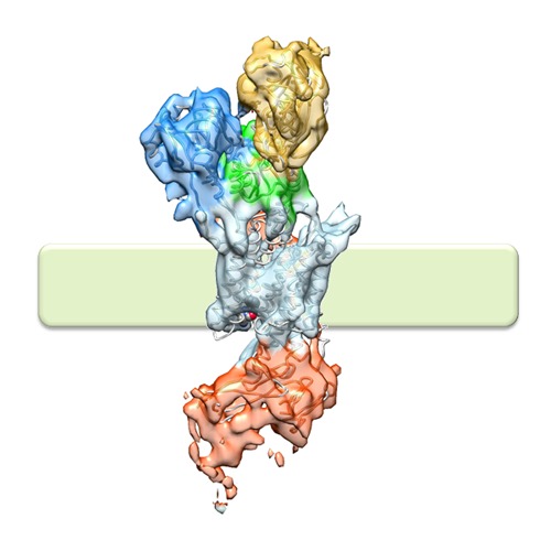
 Downloads & links
 http://ftp.pdbj.org/pub/emdb/structures/EMD-2759
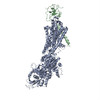


 Z
Z Y
Y X
X


 Sample components
 Processing
 Electron microscopy