Movie
Movie Controller


+ Open data

- Basic information
| Entry | Database: EMDB / ID: EMD-2692 | |||||||||
|---|---|---|---|---|---|---|---|---|---|---|
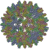
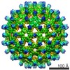
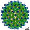
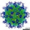
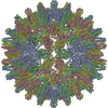
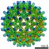
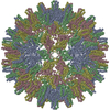
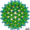
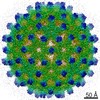
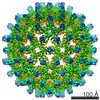
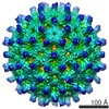
| Title | cryo-EM structure of Woodchuck Hepatitis Virus capsid | |||||||||
 Map data Map data | Cryo-EM 3-D reconstruction of Woodchuck Hepatitis Virus capsid | |||||||||
 Sample Sample |
| |||||||||
 Keywords Keywords |  Woodchuck hepatitis virus / hepatitis B virus / cryo-EM / core protein Woodchuck hepatitis virus / hepatitis B virus / cryo-EM / core protein | |||||||||
| Biological species |  Woodchuck hepatitis virus Woodchuck hepatitis virus | |||||||||
| Method | single particle reconstruction / cryo EM / Resolution: 9.7 Å | |||||||||
 Authors Authors | Kukreja A / Wang Joesph C-Y / Pierson E / Keifer DZ / Dragnea B / Jarrold MF / Zlotnick A | |||||||||
 Citation Citation | Journal: J Virol / Year: 2014 Title: Structurally similar woodchuck and human hepadnavirus core proteins have distinctly different temperature dependences of assembly. Authors: Alexander A Kukreja / Joseph C-Y Wang / Elizabeth Pierson / David Z Keifer / Lisa Selzer / Zhenning Tan / Bogdan Dragnea / Martin F Jarrold / Adam Zlotnick /  Abstract: Woodchuck hepatitis virus (WHV), a close relative of human hepatitis B virus (HBV), has been a key model for disease progression and clinical studies. Sequences of the assembly domain of WHV and HBV ...Woodchuck hepatitis virus (WHV), a close relative of human hepatitis B virus (HBV), has been a key model for disease progression and clinical studies. Sequences of the assembly domain of WHV and HBV core proteins (wCp149 and hCp149, respectively) have 65% identity, suggesting similar assembly behaviors. We report a cryo-electron microscopy (cryo-EM) structure of the WHV capsid at nanometer resolution and characterization of wCp149 assembly. At this resolution, the T=4 capsid structures of WHV and HBV are practically identical. In contrast to their structural similarity, wCp149 demonstrates enhanced assembly kinetics and stronger dimer-dimer interactions than hCp149: at 23 °C and at 100 mM ionic strength, the pseudocritical concentrations of assembly of wCp149 and hCp149 are 1.8 μM and 43.3 μM, respectively. Transmission electron microscopy reveals that wCp149 assembles into predominantly T=4 capsids with a sizeable population of larger, nonicosahedral structures. Charge detection mass spectrometry indicates that T=3 particles are extremely rare compared to the ∼ 5% observed in hCp149 reactions. Unlike hCp149, wCp149 capsid assembly is favorable over a temperature range of 4 °C to 37 °C; van't Hoff analyses relate the differences in temperature dependence to the high positive values for heat capacity, enthalpy, and entropy of wCp149 assembly. Because the final capsids are so similar, these findings suggest that free wCp149 and hCp149 undergo different structural transitions leading to assembly. The difference in the temperature dependence of wCp149 assembly may be related to the temperature range of its hibernating host. IMPORTANCE: In this paper, we present a cryo-EM structure of a WHV capsid showing its similarity to HBV. We then observe that the assembly properties of the two homologous proteins are very different. ...IMPORTANCE: In this paper, we present a cryo-EM structure of a WHV capsid showing its similarity to HBV. We then observe that the assembly properties of the two homologous proteins are very different. Unlike human HBV, the capsid protein of WHV has evolved to function in a nonhomeostatic environment. These studies yield insight into the interplay between core protein self-assembly and the host environment, which may be particularly relevant to plant viruses and viruses with zoonotic cycles involving insect vectors. | |||||||||
| History |
|
- Structure visualization
| Movie |
 Movie viewer Movie viewer |
|---|---|
| Structure viewer | EM map: SurfViewMolmilJmol/JSmol |
| Supplemental images |


- Downloads & links
-EMDB archive
| Map data | emd_2692.map.gz | 39.4 MB | EMDB map data format | |
|---|---|---|---|---|
| Header (meta data) | emd-2692-v30.xmlemd-2692.xml | 11.3 KB 11.3 KB | Display Display | EMDB header |
| Images | 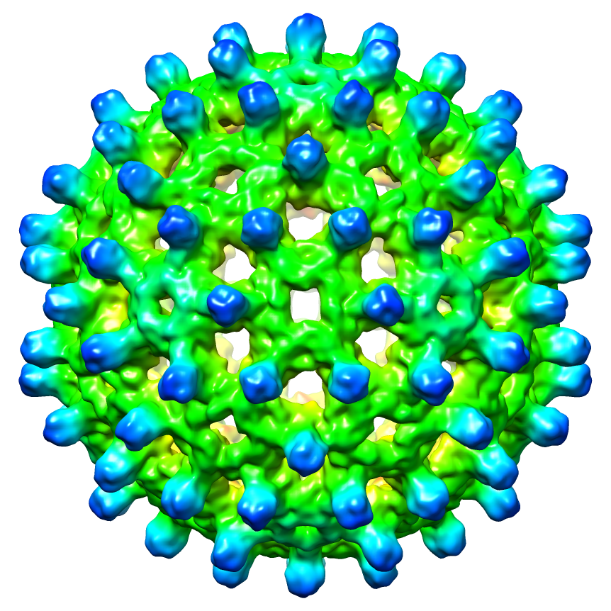 EMD-2692.png EMD-2692.png emd_2692.png emd_2692.png | 655.5 KB 655.5 KB | ||
| Archive directory |  http://ftp.pdbj.org/pub/emdb/structures/EMD-2692ftp://ftp.pdbj.org/pub/emdb/structures/EMD-2692 http://ftp.pdbj.org/pub/emdb/structures/EMD-2692ftp://ftp.pdbj.org/pub/emdb/structures/EMD-2692 | HTTPS FTP |
-Related structure data
| Similar structure data |
|---|
-Links
| EMDB pages | EMDB (EBI/PDBe) / EMDataResource |
|---|
-Map
| File | Download / File: emd_2692.map.gz / Format: CCP4 / Size: 101.6 MB / Type: IMAGE STORED AS FLOATING POINT NUMBER (4 BYTES) | ||||||||||||||||||||||||||||||||||||||||||||||||||||||||||||||||||||
|---|---|---|---|---|---|---|---|---|---|---|---|---|---|---|---|---|---|---|---|---|---|---|---|---|---|---|---|---|---|---|---|---|---|---|---|---|---|---|---|---|---|---|---|---|---|---|---|---|---|---|---|---|---|---|---|---|---|---|---|---|---|---|---|---|---|---|---|---|---|
| Annotation | Cryo-EM 3-D reconstruction of Woodchuck Hepatitis Virus capsid | ||||||||||||||||||||||||||||||||||||||||||||||||||||||||||||||||||||
| Voxel size | X=Y=Z: 1.484 Å | ||||||||||||||||||||||||||||||||||||||||||||||||||||||||||||||||||||
| Density |
| ||||||||||||||||||||||||||||||||||||||||||||||||||||||||||||||||||||
| Symmetry | Space group: 1 | ||||||||||||||||||||||||||||||||||||||||||||||||||||||||||||||||||||
| Details | EMDB XML:
CCP4 map header:
| ||||||||||||||||||||||||||||||||||||||||||||||||||||||||||||||||||||
-Supplemental data
- Sample components
-Entire : Woodchuck Hepatitis Virus core protein (wCp149)
| Entire | Name: Woodchuck Hepatitis Virus core protein (wCp149) |
|---|---|
| Components |
|
-Supramolecule #1000: Woodchuck Hepatitis Virus core protein (wCp149)
| Supramolecule | Name: Woodchuck Hepatitis Virus core protein (wCp149) / type: sample / ID: 1000 / Oligomeric state: icosahedron / Number unique components: 1 |
|---|---|
| Molecular weight | Theoretical: 4.1 MDa |
-Supramolecule #1: Woodchuck hepatitis virus
| Supramolecule | Name: Woodchuck hepatitis virus / type: virus / ID: 1 Details: WHV is an enveloped virus, but we present the truncated capsid structure here NCBI-ID: 35269 / Sci species name: Woodchuck hepatitis virus / Virus type: VIRUS-LIKE PARTICLE / Virus isolate: SPECIES / Virus enveloped: Yes / Virus empty: Yes |
|---|---|
| Host (natural) | Organism:  Marmota monax (woodchuck) / synonym: VERTEBRATES Marmota monax (woodchuck) / synonym: VERTEBRATES |
| Host system | Organism:  Escherichia coli (E. coli) / Recombinant strain: BL21 / Recombinant cell: E. coli cells / Recombinant plasmid: pET11c Escherichia coli (E. coli) / Recombinant strain: BL21 / Recombinant cell: E. coli cells / Recombinant plasmid: pET11c |
| Molecular weight | Experimental: 4.1 MDa / Theoretical: 4.1 MDa |
| Virus shell | Shell ID: 1 / Name: capsid / T number (triangulation number): 4 |
-Experimental details
-Structure determination
| Method | cryo EM |
|---|---|
 Processing Processing | single particle reconstruction |
| Aggregation state | particle |
-Sample preparation
| Concentration | 0.5 mg/mL |
|---|---|
| Buffer | pH: 7.5 / Details: 50 mM NaCl, 10 mM HEPES |
| Grid | Details: Quantifoil R2/2 |
| Vitrification | Cryogen name: ETHANE / Chamber humidity: 100 % / Instrument: FEI VITROBOT MARK III / Method: Blot for 4 seconds before plunging |
- Electron microscopy
| Microscope | JEOL 3200FS |
|---|---|
| Electron beam | Acceleration voltage: 300 kV / Electron source: FIELD EMISSION GUN |
| Electron optics | Illumination mode: FLOOD BEAM / Imaging mode: BRIGHT FIELDBright-field microscopy / Cs: 1.1 mm / Nominal defocus max: 0.0033 µm / Nominal defocus min: 0.0011 µm / Nominal magnification: 80000 |
| Specialist optics | Energy filter - Name: Omega / Energy filter - Lower energy threshold: 0.0 eV / Energy filter - Upper energy threshold: 20.0 eV |
| Sample stage | Specimen holder: 626 / Specimen holder model: GATAN LIQUID NITROGEN |
| Temperature | Average: 97.15 K |
| Alignment procedure | Legacy - Astigmatism: Objective lens astigmatism was corrected at 80,000 times magnification |
| Details | Parallel weak beam illumination |
| Date | Dec 6, 2012 |
| Image recording | Category: CCD / Film or detector model: GATAN ULTRASCAN 4000 (4k x 4k) / Digitization - Sampling interval: 1.484 µm / Number real images: 133 / Average electron dose: 25 e/Å2 / Camera length: 250 / Details: Image is recorded on CCD |
-Image processing
| CTF correction | Details: Each particle |
|---|---|
| Final angle assignment | Details: AUTO3DEM delta angle 0.7 |
| Final reconstruction | Applied symmetry - Point group: I (icosahedral) / Algorithm: OTHER / Resolution.type: BY AUTHOR / Resolution: 9.7 Å / Resolution method: OTHER / Software - Name: AUTO3DEM / Number images used: 2254 |
| Details | Particles were boxed using e2boxer.py, defocus level was measured by CTFFIND3 and image processing was done by AUTO3DEM |
-Atomic model buiding 1
| Initial model | PDB ID: Chain - #0 - Chain ID: A / Chain - #1 - Chain ID: B / Chain - #2 - Chain ID: C / Chain - #3 - Chain ID: D |
|---|---|
| Software | Name: Chimera |
| Refinement | Space: REAL / Protocol: RIGID BODY FIT |