Movie
Movie Controller

[English] 日本語
 Yorodumi
- EMDB-2598: Cryo-EM of a termination/pre-recycling complex with eRF1 and ABCE1 -
+ Open data

- Basic information
| Entry | Database: EMDB / ID: EMD-2598 | |||||||||
|---|---|---|---|---|---|---|---|---|---|---|
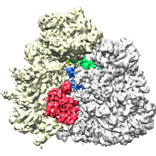
| Title | Cryo-EM of a termination/pre-recycling complex with eRF1 and ABCE1 | |||||||||
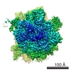
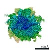
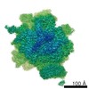
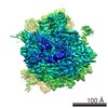
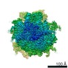
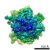
 Map data Map data | "CMV-stalled" wheat germ 80S-RNC bound to eRF1 and ABCE1. | |||||||||
 Sample Sample |
| |||||||||
 Keywords Keywords |  translation / termination / recycling / cryo-EM translation / termination / recycling / cryo-EM | |||||||||
| Function / homology |  Function and homology information Function and homology informationEukaryotic Translation Termination / cytoplasmic translational termination / translation release factor complex / translation release factor activity / ribosome disassembly / translation release factor activity, codon specific / sequence-specific mRNA binding / aminoacyl-tRNA hydrolase activity / ribosomal subunit export from nucleus / Nonsense Mediated Decay (NMD) independent of the Exon Junction Complex (EJC) ...Eukaryotic Translation Termination / cytoplasmic translational termination / translation release factor complex / translation release factor activity / ribosome disassembly / translation release factor activity, codon specific / sequence-specific mRNA binding / aminoacyl-tRNA hydrolase activity / ribosomal subunit export from nucleus / Nonsense Mediated Decay (NMD) independent of the Exon Junction Complex (EJC) / Nonsense Mediated Decay (NMD) enhanced by the Exon Junction Complex (EJC) / ribosomal small subunit binding / translational termination / ribosomal large subunit biogenesis / translational initiation / translation initiation factor activity / positive regulation of translation / DNA-templated transcription termination / rRNA processing / cytoplasmic stress granule / iron ion binding / ATP hydrolysis activity / ATP binding / nucleus / cytosol / cytoplasmSimilarity search - Function | |||||||||
| Biological species |  Triticum aestivum (bread wheat) / Triticum aestivum (bread wheat) /  Saccharomyces cerevisiae (brewer's yeast) Saccharomyces cerevisiae (brewer's yeast) | |||||||||
| Method | single particle reconstruction / cryo EM / negative staining / Resolution: 8.75 Å | |||||||||
 Authors Authors | Preis A / Heuer A / Barrio-Garcia C / Hauser A / Eyler D / Berninghausen O / Green R / Becker T / Beckmann R | |||||||||
 Citation Citation | Journal: Cell Rep / Year: 2014 Title: Cryoelectron microscopic structures of eukaryotic translation termination complexes containing eRF1-eRF3 or eRF1-ABCE1. Authors: Anne Preis / Andre Heuer / Clara Barrio-Garcia / Andreas Hauser / Daniel E Eyler / Otto Berninghausen / Rachel Green / Thomas Becker / Roland Beckmann /   Abstract: Termination and ribosome recycling are essential processes in translation. In eukaryotes, a stop codon in the ribosomal A site is decoded by a ternary complex consisting of release factors eRF1 and ...Termination and ribosome recycling are essential processes in translation. In eukaryotes, a stop codon in the ribosomal A site is decoded by a ternary complex consisting of release factors eRF1 and guanosine triphosphate (GTP)-bound eRF3. After GTP hydrolysis, eRF3 dissociates, and ABCE1 can bind to eRF1-loaded ribosomes to stimulate peptide release and ribosomal subunit dissociation. Here, we present cryoelectron microscopic (cryo-EM) structures of a pretermination complex containing eRF1-eRF3 and a termination/prerecycling complex containing eRF1-ABCE1. eRF1 undergoes drastic conformational changes: its central domain harboring the catalytically important GGQ loop is either packed against eRF3 or swung toward the peptidyl transferase center when bound to ABCE1. Additionally, in complex with eRF3, the N-terminal domain of eRF1 positions the conserved NIKS motif proximal to the stop codon, supporting its suggested role in decoding, yet it appears to be delocalized in the presence of ABCE1. These results suggest that stop codon decoding and peptide release can be uncoupled during termination. | |||||||||
| History |
|
- Structure visualization
| Movie |
Movie viewer |
|---|---|
| Structure viewer | EM map: SurfViewMolmilJmol/JSmol |
| Supplemental images |
- Downloads & links
-EMDB archive
| Map data | emd_2598.map.gz | 24.8 MB | EMDB map data format | |
|---|---|---|---|---|
| Header (meta data) | emd-2598-v30.xmlemd-2598.xml | 12.1 KB 12.1 KB | Display Display | EMDB header |
| Images |  EMD-2598-Rli_overview_side.png EMD-2598-Rli_overview_side.png | 268.6 KB | ||
| Archive directory |  http://ftp.pdbj.org/pub/emdb/structures/EMD-2598ftp://ftp.pdbj.org/pub/emdb/structures/EMD-2598 http://ftp.pdbj.org/pub/emdb/structures/EMD-2598ftp://ftp.pdbj.org/pub/emdb/structures/EMD-2598 | HTTPS FTP |
-Related structure data
| Related structure data | 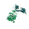 4crmMC 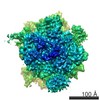 2597C  4crnC M: atomic model generated by this map C: citing same article ( |
|---|---|
| Similar structure data |
-Links
| EMDB pages | EMDB (EBI/PDBe) / EMDataResource |
|---|---|
| Related items in Molecule of the Month |


-Map
| File | Download / File: emd_2598.map.gz / Format: CCP4 / Size: 185.7 MB / Type: IMAGE STORED AS FLOATING POINT NUMBER (4 BYTES) | ||||||||||||||||||||||||||||||||||||||||||||||||||||||||||||||||||||
|---|---|---|---|---|---|---|---|---|---|---|---|---|---|---|---|---|---|---|---|---|---|---|---|---|---|---|---|---|---|---|---|---|---|---|---|---|---|---|---|---|---|---|---|---|---|---|---|---|---|---|---|---|---|---|---|---|---|---|---|---|---|---|---|---|---|---|---|---|---|
| Annotation | "CMV-stalled" wheat germ 80S-RNC bound to eRF1 and ABCE1. | ||||||||||||||||||||||||||||||||||||||||||||||||||||||||||||||||||||
| Voxel size | X=Y=Z: 1.2375 Å | ||||||||||||||||||||||||||||||||||||||||||||||||||||||||||||||||||||
| Density |
| ||||||||||||||||||||||||||||||||||||||||||||||||||||||||||||||||||||
| Symmetry | Space group: 1 | ||||||||||||||||||||||||||||||||||||||||||||||||||||||||||||||||||||
| Details | EMDB XML:
CCP4 map header:
| ||||||||||||||||||||||||||||||||||||||||||||||||||||||||||||||||||||
-Supplemental data
- Sample components
-Entire : "CMV"-stalled wheat germ 80S-RNC bound to eRF1 and ABCE1-ADPNP
| Entire | Name: "CMV"-stalled wheat germ 80S-RNC bound to eRF1 and ABCE1-ADPNP |
|---|---|
| Components |
|
-Supramolecule #1000: "CMV"-stalled wheat germ 80S-RNC bound to eRF1 and ABCE1-ADPNP
| Supramolecule | Name: "CMV"-stalled wheat germ 80S-RNC bound to eRF1 and ABCE1-ADPNP type: sample / ID: 1000 Oligomeric state: One ribosome binds to one eRF1 and one ABCE1 Number unique components: 3 |
|---|
-Supramolecule #1: 80S ribosome
| Supramolecule | Name: 80S ribosome / type: complex / ID: 1 / Recombinant expression: No / Ribosome-details: ribosome-eukaryote: ALL |
|---|---|
| Source (natural) | Organism: Triticum aestivum (bread wheat) / synonym: Bread wheat / Tissue: seed |
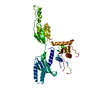
-Macromolecule #1: Sup45
| Macromolecule | Name: Sup45 / type: protein_or_peptide / ID: 1 / Name.synonym: eRF1 / Number of copies: 1 / Oligomeric state: 1 / Recombinant expression: Yes |
|---|---|
| Source (natural) | Organism: Saccharomyces cerevisiae (brewer's yeast) / synonym: Baker's Yeast / Location in cell: cytoplasm |
| Molecular weight | Theoretical: 49 KDa |
| Recombinant expression | Organism:  Escherichia coli (E. coli) / Recombinant plasmid: pTYB2 Escherichia coli (E. coli) / Recombinant plasmid: pTYB2 |
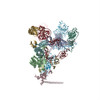
-Macromolecule #2: Rli1
| Macromolecule | Name: Rli1 / type: protein_or_peptide / ID: 2 / Name.synonym: ABCE1 / Number of copies: 1 / Oligomeric state: 1 / Recombinant expression: Yes |
|---|---|
| Source (natural) | Organism: Saccharomyces cerevisiae (brewer's yeast) / synonym: Baker's yeast / Location in cell: cytoplasm |
| Molecular weight | Theoretical: 68 KDa |
| Recombinant expression | Organism: Saccharomyces cerevisiae (brewer's yeast) / Recombinant plasmid: pYES2 |
-Experimental details
-Structure determination
| Method | negative staining, cryo EM |
|---|---|
 Processing Processing | single particle reconstruction |
| Aggregation state | particle |
-Sample preparation
| Buffer | pH: 7.5 Details: 20 mM HEPES pH 7.5, 200 mM KCl, 1.5 MgCl2, 2 mM DTT, 0.01 mg/ml cycloheximide, 0.05 % Nikkol, 0.03 % DBC, 0.5 mM ADPNP). |
|---|---|
| Staining | Type: NEGATIVE / Details: Cryo-EM |
| Grid | Details: Sec61 was added at a five-fold molar excess to saturate the hydrophobic signal-anchor sequence and avoid orientational bias on the cryo-grids |
| Vitrification | Cryogen name: ETHANE / Chamber humidity: 100 % / Instrument: FEI VITROBOT MARK IV / Method: Blot for 3 seconds before plunging |
- Electron microscopy
| Microscope | FEI TITAN KRIOS |
|---|---|
| Electron beam | Acceleration voltage: 200 kV / Electron source: FIELD EMISSION GUN |
| Electron optics | Calibrated magnification: 147136 / Illumination mode: FLOOD BEAM / Imaging mode: BRIGHT FIELDBright-field microscopy / Cs: 2.7 mm |
| Sample stage | Specimen holder model: FEI TITAN KRIOS AUTOGRID HOLDER |
| Date | Feb 20, 2013 |
| Image recording | Category: CCD / Film or detector model: TVIPS TEMCAM-F416 (4k x 4k) / Digitization - Sampling interval: 15.6 µm / Average electron dose: 20 e/Å2 / Bits/pixel: 16 |
| Experimental equipment |  Model: Titan Krios / Image courtesy: FEI Company |
-Image processing
| CTF correction | Details: on volumes (SPIDER) |
|---|---|
| Final reconstruction | Applied symmetry - Point group: C1 (asymmetric) / Algorithm: OTHER / Resolution.type: BY AUTHOR / Resolution: 8.75 Å / Resolution method: OTHER / Software - Name: StarFish, SPIDER / Details: Data-subset resulted from computational sorting / Number images used: 39309 |
| Details | The particles were picked with starfish_boxing version 0.2.0, which is part of the new StarFish single particle analysis program suite. |
-Atomic model buiding 1
| Initial model | PDB ID: |
|---|---|
| Software | Name: Chimera, Coot, MDFF |
| Details | A homology model of eRF1 was created using HHPred. The domains were separately fitted by manual docking using program Coot. |
| Refinement | Space: REAL / Protocol: RIGID BODY FIT |
| Output model | PDB-4crm: |