 Movie
Movie Controller

[English] 日本語
 Yorodumi
- EMDB-2581: The structures of cytosolic and plastid-located glutamine synthet... -
+ Open data

- Basic information
| Entry | Database: EMDB / ID: EMD-2581 | |||||||||
|---|---|---|---|---|---|---|---|---|---|---|
| Title | The structures of cytosolic and plastid-located glutamine synthetases from Medicago truncatula reveal a common and dynamic architecture | |||||||||
 Map data Map data | gsii-a reconstruction from M. trucantula | |||||||||
 Sample Sample |
| |||||||||
 Keywords Keywords | glutamine synthetases / Medicago trucantula | |||||||||
| Biological species |   Medicago truncatula (barrel medic) Medicago truncatula (barrel medic) | |||||||||
| Method | single particle reconstruction / cryo EM / Resolution: 20.0 Å | |||||||||
 Authors Authors | Torreira E / Seabra AR / Marriott H / Zhou M / Llorca O / Robinson CV / Carvalho HG / Fernandez-Tornero C / Pereira PJB | |||||||||
 Citation Citation | Journal: Acta Crystallogr D Biol Crystallogr / Year: 2014 Title: The structures of cytosolic and plastid-located glutamine synthetases from Medicago truncatula reveal a common and dynamic architecture. Authors: Eva Torreira / Ana Rita Seabra / Hazel Marriott / Min Zhou / Óscar Llorca / Carol V Robinson / Helena G Carvalho / Carlos Fernández-Tornero / Pedro José Barbosa Pereira /    Abstract: The first step of nitrogen assimilation in higher plants, the energy-driven incorporation of ammonia into glutamate, is catalyzed by glutamine synthetase. This central process yields the readily ...The first step of nitrogen assimilation in higher plants, the energy-driven incorporation of ammonia into glutamate, is catalyzed by glutamine synthetase. This central process yields the readily metabolizable glutamine, which in turn is at the basis of all subsequent biosynthesis of nitrogenous compounds. The essential role performed by glutamine synthetase makes it a prime target for herbicidal compounds, but also a suitable intervention point for the improvement of crop yields. Although the majority of crop plants are dicotyledonous, little is known about the structural organization of glutamine synthetase in these organisms and about the functional differences between the different isoforms. Here, the structural characterization of two glutamine synthetase isoforms from the model legume Medicago truncatula is reported: the crystallographic structure of cytoplasmic GSII-1a and an electron cryomicroscopy reconstruction of plastid-located GSII-2a. Together, these structural models unveil a decameric organization of dicotyledonous glutamine synthetase, with two pentameric rings weakly connected by inter-ring loops. Moreover, rearrangement of these dynamic loops changes the relative orientation of the rings, suggesting a zipper-like mechanism for their assembly into a decameric enzyme. Finally, the atomic structure of M. truncatula GSII-1a provides important insights into the structural determinants of herbicide resistance in this family of enzymes, opening new avenues for the development of herbicide-resistant plants. | |||||||||
| History |
|
- Structure visualization
| Movie |
 Movie viewer Movie viewer |
|---|---|
| Structure viewer | EM map: SurfViewMolmilJmol/JSmol |
| Supplemental images |

- Downloads & links
-EMDB archive
| Map data | emd_2581.map.gz | 1.5 MB | EMDB map data format | |
|---|---|---|---|---|
| Header (meta data) | emd-2581-v30.xmlemd-2581.xml | 9.3 KB 9.3 KB | Display Display | EMDB header |
| Images | 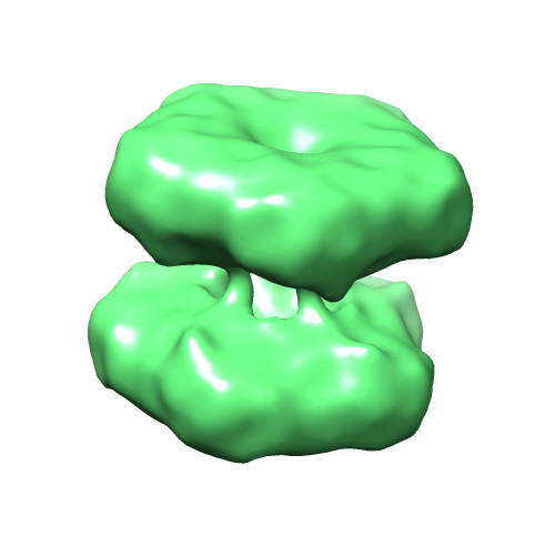 emd_2581.png emd_2581.png | 84.5 KB | ||
| Archive directory |  http://ftp.pdbj.org/pub/emdb/structures/EMD-2581ftp://ftp.pdbj.org/pub/emdb/structures/EMD-2581 http://ftp.pdbj.org/pub/emdb/structures/EMD-2581ftp://ftp.pdbj.org/pub/emdb/structures/EMD-2581 | HTTPS FTP |
-Related structure data


-Links
| EMDB pages | EMDB (EBI/PDBe) / EMDataResource |
|---|
-Map
| File | Download / File: emd_2581.map.gz / Format: CCP4 / Size: 20.3 MB / Type: IMAGE STORED AS FLOATING POINT NUMBER (4 BYTES) | ||||||||||||||||||||||||||||||||||||||||||||||||||||||||||||||||||||
|---|---|---|---|---|---|---|---|---|---|---|---|---|---|---|---|---|---|---|---|---|---|---|---|---|---|---|---|---|---|---|---|---|---|---|---|---|---|---|---|---|---|---|---|---|---|---|---|---|---|---|---|---|---|---|---|---|---|---|---|---|---|---|---|---|---|---|---|---|---|
| Annotation | gsii-a reconstruction from M. trucantula | ||||||||||||||||||||||||||||||||||||||||||||||||||||||||||||||||||||
| Voxel size | X=Y=Z: 1.62 Å | ||||||||||||||||||||||||||||||||||||||||||||||||||||||||||||||||||||
| Density |
| ||||||||||||||||||||||||||||||||||||||||||||||||||||||||||||||||||||
| Symmetry | Space group: 1 | ||||||||||||||||||||||||||||||||||||||||||||||||||||||||||||||||||||
| Details | EMDB XML:
CCP4 map header:
| ||||||||||||||||||||||||||||||||||||||||||||||||||||||||||||||||||||
-Supplemental data
- Sample components
-Entire : gsii-a from M. trucantula
| Entire | Name: gsii-a from M. trucantula |
|---|---|
| Components |
|
-Supramolecule #1000: gsii-a from M. trucantula
| Supramolecule | Name: gsii-a from M. trucantula / type: sample / ID: 1000 / Oligomeric state: 2-pentameric rings / Number unique components: 1 |
|---|---|
| Molecular weight | Experimental: 441 KDa / Theoretical: 440 KDa / Method: Native mass-spec |
-Macromolecule #1: MtGSII-2a
| Macromolecule | Name: MtGSII-2a / type: protein_or_peptide / ID: 1 / Number of copies: 10 / Oligomeric state: Decamer / Recombinant expression: Yes |
|---|---|
| Source (natural) | Organism: Medicago truncatula (barrel medic) / synonym: Barrel medic / Location in cell: plastid |
| Molecular weight | Experimental: 441 KDa / Theoretical: 440 KDa |
| Recombinant expression | Organism:  Escherichia coli BL21(DE3) (bacteria) / Recombinant strain: CodonPlus RIL / Recombinant plasmid: pET-24-d(+) Escherichia coli BL21(DE3) (bacteria) / Recombinant strain: CodonPlus RIL / Recombinant plasmid: pET-24-d(+) |
-Experimental details
-Structure determination
| Method | cryo EM |
|---|---|
 Processing Processing | single particle reconstruction |
| Aggregation state | particle |
-Sample preparation
| Concentration | 0.3 mg/mL |
|---|---|
| Buffer | pH: 7.4 / Details: 10 mM HEPES PH 7.4 |
| Grid | Details: Glow-discharged QUANTIFOIL R 2/2 holey grids |
| Vitrification | Cryogen name: ETHANE / Instrument: GATAN CRYOPLUNGE 3 |
- Electron microscopy
| Microscope | JEOL 2200FS |
|---|---|
| Electron beam | Acceleration voltage: 200 kV / Electron source: FIELD EMISSION GUN |
| Electron optics | Calibrated magnification: 83393 / Illumination mode: FLOOD BEAM / Imaging mode: BRIGHT FIELDBright-field microscopy |
| Sample stage | Specimen holder model: GATAN LIQUID NITROGEN |
| Date | Nov 1, 2012 |
| Image recording | Category: CCD / Film or detector model: GATAN ULTRASCAN 4000 (4k x 4k) |
| Tilt angle min | 0 |
| Tilt angle max | 0 |
-Image processing
| CTF correction | Details: CTFFIND and BSOFT |
|---|---|
| Final reconstruction | Applied symmetry - Point group: D5 (2x5 fold dihedral) / Algorithm: OTHER / Resolution.type: BY AUTHOR / Resolution: 20.0 Å / Resolution method: OTHER / Software - Name: EMAN1.9, Xmipp / Number images used: 3117 |
| Details | The final reconstruction was determined employing just perfect side-views. |
-Atomic model buiding 1
| Initial model | PDB ID: |
|---|---|
| Software | Name: Chimera |
| Refinement | Space: REAL / Protocol: RIGID BODY FIT |
