 Movie
Movie Controller


+ Open data

- Basic information
| Entry | Database: EMDB / ID: EMD-2572 | |||||||||
|---|---|---|---|---|---|---|---|---|---|---|
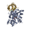
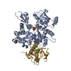
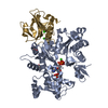
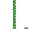
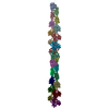
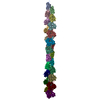
| Title | Cryo-EM structure of Plasmodium falciparum actin I | |||||||||
 Map data Map data | Cryo-EM structure of Plasmodium falciparum actin I | |||||||||
 Sample Sample |
| |||||||||
 Keywords Keywords | Plasmodium falciparum Actin I /  malaria parasite malaria parasite | |||||||||
| Function / homology |  Function and homology information Function and homology informationActins signature 1. / Actin, conserved site / Actins signature 2. / Actin/actin-like conserved site / Actins and actin-related proteins signature. / Actin / Actin family / Actin / ATPase, nucleotide binding domainSimilarity search - Domain/homology | |||||||||
| Biological species |  Plasmodium falciparum (malaria parasite P. falciparum) Plasmodium falciparum (malaria parasite P. falciparum) | |||||||||
| Method | single particle reconstruction / cryo EM / Resolution: 25.0 Å | |||||||||
 Authors Authors | Vahokoski J / Bhargav SP / Desfosses A / Andreadaki M / Kumpula E / Ignatev A / Martinez SM / Lepper S / Frischknecht F / Siden-Kiamos I ...Vahokoski J / Bhargav SP / Desfosses A / Andreadaki M / Kumpula E / Ignatev A / Martinez SM / Lepper S / Frischknecht F / Siden-Kiamos I / Sachse C / Kursula I | |||||||||
 Citation Citation | Journal: PLoS Pathog / Year: 2014 Title: Structural differences explain diverse functions of Plasmodium actins. Authors: Juha Vahokoski / Saligram Prabhakar Bhargav / Ambroise Desfosses / Maria Andreadaki / Esa-Pekka Kumpula / Silvia Muñico Martinez / Alexander Ignatev / Simone Lepper / Friedrich Frischknecht ...Authors: Juha Vahokoski / Saligram Prabhakar Bhargav / Ambroise Desfosses / Maria Andreadaki / Esa-Pekka Kumpula / Silvia Muñico Martinez / Alexander Ignatev / Simone Lepper / Friedrich Frischknecht / Inga Sidén-Kiamos / Carsten Sachse / Inari Kursula /    Abstract: Actins are highly conserved proteins and key players in central processes in all eukaryotic cells. The two actins of the malaria parasite are among the most divergent eukaryotic actins and also ...Actins are highly conserved proteins and key players in central processes in all eukaryotic cells. The two actins of the malaria parasite are among the most divergent eukaryotic actins and also differ from each other more than isoforms in any other species. Microfilaments have not been directly observed in Plasmodium and are presumed to be short and highly dynamic. We show that actin I cannot complement actin II in male gametogenesis, suggesting critical structural differences. Cryo-EM reveals that Plasmodium actin I has a unique filament structure, whereas actin II filaments resemble canonical F-actin. Both Plasmodium actins hydrolyze ATP more efficiently than α-actin, and unlike any other actin, both parasite actins rapidly form short oligomers induced by ADP. Crystal structures of both isoforms pinpoint several structural changes in the monomers causing the unique polymerization properties. Inserting the canonical D-loop to Plasmodium actin I leads to the formation of long filaments in vitro. In vivo, this chimera restores gametogenesis in parasites lacking actin II, suggesting that stable filaments are required for exflagellation. Together, these data underline the divergence of eukaryotic actins and demonstrate how structural differences in the monomers translate into filaments with different properties, implying that even eukaryotic actins have faced different evolutionary pressures and followed different paths for developing their polymerization properties. | |||||||||
| History |
|
- Structure visualization
| Movie |
Movie viewer |
|---|---|
| Structure viewer | EM map: SurfViewMolmilJmol/JSmol |
| Supplemental images |

- Downloads & links
-EMDB archive
| Map data | emd_2572.map.gz | 632.7 KB | EMDB map data format | |
|---|---|---|---|---|
| Header (meta data) | emd-2572-v30.xmlemd-2572.xml | 9.9 KB 9.9 KB | Display Display | EMDB header |
| Images | 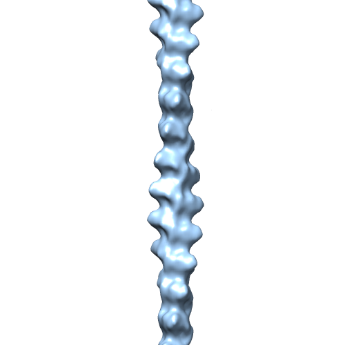 EMD-2572-Actin-I_for-EMDB.png EMD-2572-Actin-I_for-EMDB.png | 51 KB | ||
| Archive directory |  http://ftp.pdbj.org/pub/emdb/structures/EMD-2572ftp://ftp.pdbj.org/pub/emdb/structures/EMD-2572 http://ftp.pdbj.org/pub/emdb/structures/EMD-2572ftp://ftp.pdbj.org/pub/emdb/structures/EMD-2572 | HTTPS FTP |
-Related structure data
-Links
| EMDB pages | EMDB (EBI/PDBe) / EMDataResource |
|---|---|
| Related items in Molecule of the Month |


-Map
| File | Download / File: emd_2572.map.gz / Format: CCP4 / Size: 917 KB / Type: IMAGE STORED AS FLOATING POINT NUMBER (4 BYTES) | ||||||||||||||||||||||||||||||||||||||||||||||||||||||||||||||||||||
|---|---|---|---|---|---|---|---|---|---|---|---|---|---|---|---|---|---|---|---|---|---|---|---|---|---|---|---|---|---|---|---|---|---|---|---|---|---|---|---|---|---|---|---|---|---|---|---|---|---|---|---|---|---|---|---|---|---|---|---|---|---|---|---|---|---|---|---|---|---|
| Annotation | Cryo-EM structure of Plasmodium falciparum actin I | ||||||||||||||||||||||||||||||||||||||||||||||||||||||||||||||||||||
| Voxel size | X=Y=Z: 4.412 Å | ||||||||||||||||||||||||||||||||||||||||||||||||||||||||||||||||||||
| Density |
| ||||||||||||||||||||||||||||||||||||||||||||||||||||||||||||||||||||
| Symmetry | Space group: 1 | ||||||||||||||||||||||||||||||||||||||||||||||||||||||||||||||||||||
| Details | EMDB XML:
CCP4 map header:
| ||||||||||||||||||||||||||||||||||||||||||||||||||||||||||||||||||||
-Supplemental data
- Sample components
-Entire : Plasmodium falciparum Actin I
| Entire | Name: Plasmodium falciparum Actin I |
|---|---|
| Components |
|
-Supramolecule #1000: Plasmodium falciparum Actin I
| Supramolecule | Name: Plasmodium falciparum Actin I / type: sample / ID: 1000 / Number unique components: 1 |
|---|
-Macromolecule #1: Plasmodium actin I
| Macromolecule | Name: Plasmodium actin I / type: protein_or_peptide / ID: 1 / Recombinant expression: Yes |
|---|---|
| Source (natural) | Organism: Plasmodium falciparum (malaria parasite P. falciparum) |
| Recombinant expression | Organism:   Spodoptera frugiperda (fall armyworm) / Recombinant strain: Sf21 Spodoptera frugiperda (fall armyworm) / Recombinant strain: Sf21 |
| Sequence | UniProtKB: Actin I |
-Experimental details
-Structure determination
| Method | cryo EM |
|---|---|
 Processing Processing | single particle reconstruction |
| Aggregation state | helical array |
-Sample preparation
| Concentration | 0.3 mg/mL |
|---|---|
| Buffer | Details: 50 mM (pH 8.0) Tris-HCl, 500 mM KCl, 20 mM MgCl2, 50 mM DTT, and 10 mM ATP, JAS 5 uM |
| Grid | Details: glow-discharged holey carbon grids (Quantifoil R 2/2) |
| Vitrification | Cryogen name: ETHANE / Chamber humidity: 70 % / Chamber temperature: 120 K / Instrument: LEICA EM GP Method: Polymerized samples were applied in 3-uL aliquots onto freshly glow-discharged holey carbon grids (Quantifoil R 2/2) at 295 K and 70% humidity and vitrified in liquid ethane using a Leica EM ...Method: Polymerized samples were applied in 3-uL aliquots onto freshly glow-discharged holey carbon grids (Quantifoil R 2/2) at 295 K and 70% humidity and vitrified in liquid ethane using a Leica EM GP vitrification robot. |
- Electron microscopy
| Microscope | FEI TECNAI F20 |
|---|---|
| Electron beam | Acceleration voltage: 200 kV / Electron source: FIELD EMISSION GUN |
| Electron optics | Calibrated magnification: 69000 / Illumination mode: FLOOD BEAM / Imaging mode: BRIGHT FIELDBright-field microscopy / Cs: 2.0 mm / Nominal defocus max: 7.0 µm / Nominal defocus min: 1.0 µm / Nominal magnification: 69000 |
| Sample stage | Specimen holder: 93 K / Specimen holder model: GATAN LIQUID NITROGEN |
| Date | Feb 6, 2013 |
| Image recording | Category: CCD / Film or detector model: GATAN ULTRASCAN 4000 (4k x 4k) / Number real images: 75 |
| Experimental equipment |  Model: Tecnai F20 / Image courtesy: FEI Company |
-Image processing
| CTF correction | Details: Each particle (convolved) |
|---|---|
| Final reconstruction | Applied symmetry - Helical parameters - Δz: 27.69 Å Applied symmetry - Helical parameters - Δ&Phi: 167.52 ° Applied symmetry - Helical parameters - Axial symmetry: C1 (asymmetric) Resolution.type: BY AUTHOR / Resolution: 25.0 Å / Resolution method: OTHER / Software - Name: SPRING / Number images used: 3513 |
| Details | For 3D structure determination, segments were excised using a regular step size of 70 Angstrom, convolved by their respective CTF and further reconstructed as described (Sachse et al. 2007) using the software SPRING (Desfosses et al. 2014) |
