 Movie
Movie Controller

[English] 日本語
 Yorodumi
- EMDB-2553: Structural analysis of the interaction between C3b complement pro... -
+ Open data

- Basic information
| Entry | Database: EMDB / ID: EMD-2553 | |||||||||
|---|---|---|---|---|---|---|---|---|---|---|
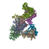
| Title | Structural analysis of the interaction between C3b complement protein and H17(Fab). | |||||||||
 Map data Map data | This is the 3D structure of C3b bound to H17 (Fab) | |||||||||
 Sample Sample |
| |||||||||
 Keywords Keywords |  Complement system / C3b / Fab / H17 antibody Complement system / C3b / Fab / H17 antibody | |||||||||
| Biological species |  Homo sapiens (human) / Homo sapiens (human) /  Mus musculus (house mouse) Mus musculus (house mouse) | |||||||||
| Method | single particle reconstruction / negative staining / Resolution: 30.0 Å | |||||||||
 Authors Authors | Paixao-Cavalcante D / Torreira E / Lindorfer MA / Rodriguez de Cordoba S / Morgan BP / Taylor RP / Llorca O / Harris CL | |||||||||
 Citation Citation | Journal: J Immunol / Year: 2014 Title: A humanized antibody that regulates the alternative pathway convertase: potential for therapy of renal disease associated with nephritic factors. Authors: Danielle Paixão-Cavalcante / Eva Torreira / Margaret A Lindorfer / Santiago Rodriguez de Cordoba / B Paul Morgan / Ronald P Taylor / Oscar Llorca / Claire L Harris /  Abstract: Dysregulation of the complement alternative pathway can cause disease in various organs that may be life-threatening. Severe alternative pathway dysregulation can be triggered by autoantibodies to ...Dysregulation of the complement alternative pathway can cause disease in various organs that may be life-threatening. Severe alternative pathway dysregulation can be triggered by autoantibodies to the C3 convertase, termed nephritic factors, which cause pathological stabilization of the convertase enzyme and confer resistance to innate control mechanisms; unregulated complement consumption followed by deposition of C3 fragments in tissues ensues. The mAb, 3E7, and its humanized derivative, H17, have been shown previously to specifically bind activated C3 and prevent binding of both the activating protein, factor B, and the inhibitor, factor H, which are opposite effects that complicate its potential for therapy. Using ligand binding assays, functional assays, and electron microscopy, we show that these Abs bind C3b via a site that overlaps the binding site on C3 for the Ba domain within factor B, thereby blocking an interaction essential for convertase formation. Both Abs also bind the preformed convertase, C3bBb, and provide powerful inhibition of complement activation by preventing cleavage of C3. Critically, the Abs also bound and inhibited C3 cleavage by the nephritic factor-stabilized convertase. We suggest that by preventing enzyme formation and/or cleavage of C3 to its active downstream fragments, H17 may be an effective therapy for conditions caused by severe dysregulation of the C3 convertase and, in particular, those that involve nephritic factors, such as dense deposit disease. | |||||||||
| History |
|
- Structure visualization
| Movie |
 Movie viewer Movie viewer |
|---|---|
| Structure viewer | EM map: SurfViewMolmilJmol/JSmol |
| Supplemental images |

- Downloads & links
-EMDB archive
| Map data | emd_2553.map.gz | 1.2 MB | EMDB map data format | |
|---|---|---|---|---|
| Header (meta data) | emd-2553-v30.xmlemd-2553.xml | 10.2 KB 10.2 KB | Display Display | EMDB header |
| Images | 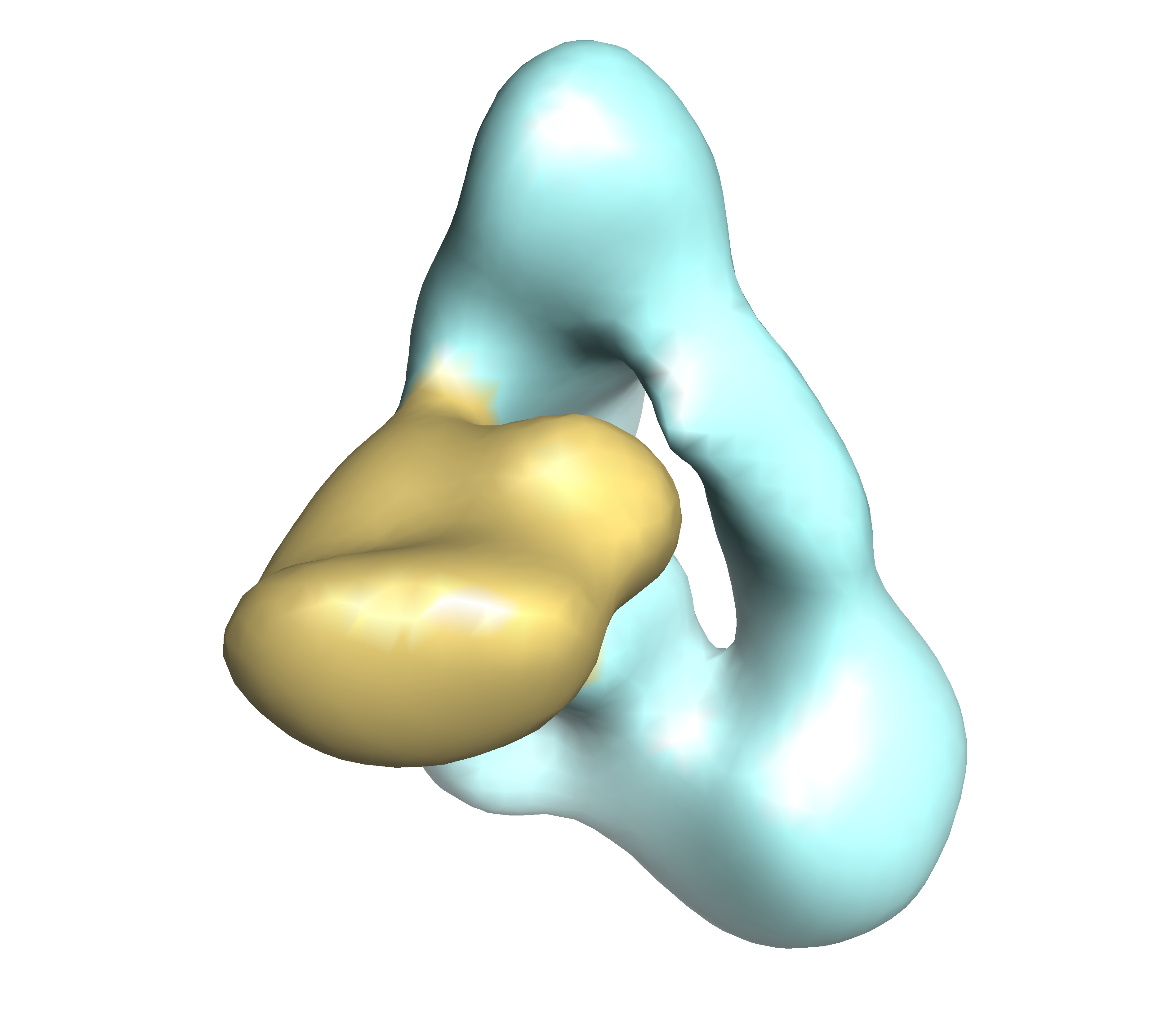 EMD-2553.jpg EMD-2553.jpg | 1.6 MB | ||
| Archive directory |  http://ftp.pdbj.org/pub/emdb/structures/EMD-2553ftp://ftp.pdbj.org/pub/emdb/structures/EMD-2553 http://ftp.pdbj.org/pub/emdb/structures/EMD-2553ftp://ftp.pdbj.org/pub/emdb/structures/EMD-2553 | HTTPS FTP |
-Related structure data
| Similar structure data |
|---|

-Links
| EMDB pages | EMDB (EBI/PDBe) / EMDataResource |
|---|
-Map
| File | Download / File: emd_2553.map.gz / Format: CCP4 / Size: 1.4 MB / Type: IMAGE STORED AS FLOATING POINT NUMBER (4 BYTES) | ||||||||||||||||||||||||||||||||||||||||||||||||||||||||||||||||||||
|---|---|---|---|---|---|---|---|---|---|---|---|---|---|---|---|---|---|---|---|---|---|---|---|---|---|---|---|---|---|---|---|---|---|---|---|---|---|---|---|---|---|---|---|---|---|---|---|---|---|---|---|---|---|---|---|---|---|---|---|---|---|---|---|---|---|---|---|---|---|
| Annotation | This is the 3D structure of C3b bound to H17 (Fab) | ||||||||||||||||||||||||||||||||||||||||||||||||||||||||||||||||||||
| Voxel size | X=Y=Z: 4.56 Å | ||||||||||||||||||||||||||||||||||||||||||||||||||||||||||||||||||||
| Density |
| ||||||||||||||||||||||||||||||||||||||||||||||||||||||||||||||||||||
| Symmetry | Space group: 1 | ||||||||||||||||||||||||||||||||||||||||||||||||||||||||||||||||||||
| Details | EMDB XML:
CCP4 map header:
| ||||||||||||||||||||||||||||||||||||||||||||||||||||||||||||||||||||
-Supplemental data
- Sample components
-Entire : C3b
| Entire | Name: C3b |
|---|---|
| Components |
|
-Supramolecule #1000: C3b
| Supramolecule | Name: C3b / type: sample / ID: 1000 Details: Sample behaves as a unique peak in gel filtration when analyzed using a Duperdex 200 column. Oligomeric state: One molecule of C3b bound to one Fab. / Number unique components: 2 |
|---|---|
| Molecular weight | Experimental: 230 KDa / Theoretical: 230 KDa / Method: SDS-PAGE |
-Macromolecule #1: C3b
| Macromolecule | Name: C3b / type: protein_or_peptide / ID: 1 / Number of copies: 1 / Oligomeric state: Monomer / Recombinant expression: No |
|---|---|
| Source (natural) | Organism: Homo sapiens (human) / synonym: Human / Location in cell: plasma |
| Molecular weight | Experimental: 180 KDa / Theoretical: 180 KDa |
-Macromolecule #2: Fab fragment of H17 antibody
| Macromolecule | Name: Fab fragment of H17 antibody / type: protein_or_peptide / ID: 2 / Number of copies: 1 / Oligomeric state: 1 / Recombinant expression: Yes |
|---|---|
| Source (natural) | Organism: Mus musculus (house mouse) / synonym: mouse / Cell: NS0 cells / Location in cell: supernatant |
| Molecular weight | Experimental: 50 KDa |
| Recombinant expression | Recombinant cell: NS0 cells (murine myeloma cell line) |
-Experimental details
-Structure determination
| Method | negative staining |
|---|---|
 Processing Processing | single particle reconstruction |
| Aggregation state | particle |
-Sample preparation
| Concentration | 0.01 mg/mL |
|---|---|
| Buffer | pH: 7.4 / Details: 25 mM Tris HCl pH 7.4, 150 mM NaCl, 10 mM DTT |
| Staining | Type: NEGATIVE Details: Grids containing adsorbed protein were floated on 2% uranyl formate for 2 minutes |
| Vitrification | Cryogen name: NONE / Instrument: OTHER |
- Electron microscopy
| Microscope | JEOL 1230 |
|---|---|
| Electron beam | Acceleration voltage: 100 kV / Electron source: TUNGSTEN HAIRPIN |
| Electron optics | Illumination mode: FLOOD BEAM / Imaging mode: BRIGHT FIELDBright-field microscopy / Cs: 2.9 mm / Nominal magnification: 50000 |
| Sample stage | Specimen holder model: JEOL |
| Date | Sep 1, 2011 |
| Image recording | Category: CCD / Film or detector model: TVIPS TEMCAM-F416 (4k x 4k) |
-Image processing
| Final reconstruction | Applied symmetry - Point group: C1 (asymmetric) / Algorithm: OTHER / Resolution.type: BY AUTHOR / Resolution: 30.0 Å / Resolution method: OTHER / Software - Name: EMAN1.9, XMIPP, 2.4 / Number images used: 7227 |
|---|
-Atomic model buiding 1
| Initial model | PDB ID: |
|---|---|
| Software | Name: Chimera |
| Refinement | Space: REAL / Protocol: RIGID BODY FIT |

-Atomic model buiding 2
| Initial model | PDB ID: Chain - #0 - Chain ID: A / Chain - #1 - Chain ID: B |
|---|---|
| Software | Name: Chimera |
| Refinement | Space: REAL / Protocol: RIGID BODY FIT |

