 Movie
Movie Controller

[English] 日本語
 Yorodumi
- EMDB-2548: Single particle electron cryomicroscopy of the complex between th... -
+ Open data

- Basic information
| Entry | Database: EMDB / ID: EMD-2548 | |||||||||
|---|---|---|---|---|---|---|---|---|---|---|
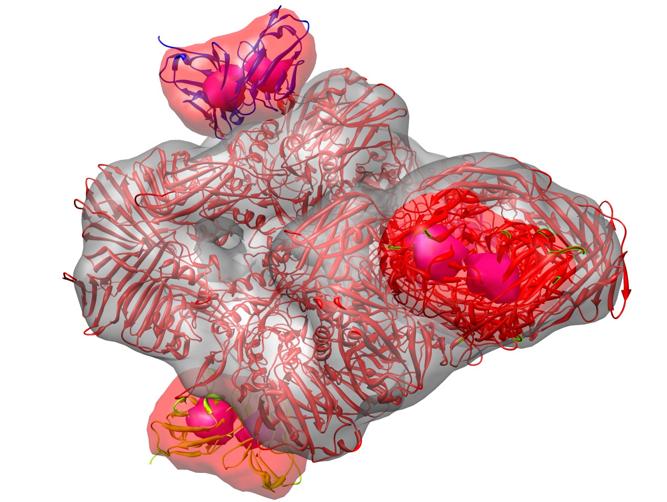
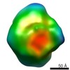
| Title | Single particle electron cryomicroscopy of the complex between the E.coli enzyme beta-galactosidase and the single chain Fv antibody scFv13R4. | |||||||||
 Map data Map data | Map of the structure of the complex between beta-galactosidase and the scFv13R4 single chain antibody domain. | |||||||||
 Sample Sample |
| |||||||||
 Keywords Keywords |  beta-galactosidase / antibody / scFv13R4 / single particle cryoEM beta-galactosidase / antibody / scFv13R4 / single particle cryoEM | |||||||||
| Function / homology |  Function and homology information Function and homology informationalkali metal ion binding / lactose catabolic process / beta-galactosidase complex / beta-galactosidase / beta-galactosidase activity / immunoglobulin complex / immunoglobulin mediated immune response / antigen binding / carbohydrate binding / adaptive immune response ...alkali metal ion binding / lactose catabolic process / beta-galactosidase complex / beta-galactosidase / beta-galactosidase activity / immunoglobulin complex / immunoglobulin mediated immune response / antigen binding / carbohydrate binding / adaptive immune response / immune response / magnesium ion binding / extracellular space / identical protein bindingSimilarity search - Function | |||||||||
| Biological species |  Escherichia coli (E. coli) / Escherichia coli (E. coli) /  Mus musculus (house mouse) Mus musculus (house mouse) | |||||||||
| Method | single particle reconstruction / cryo EM / negative staining / Resolution: 13.0 Å | |||||||||
 Authors Authors | Vinothkumar KR / McMullan G / Henderson R | |||||||||
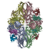
 Citation Citation | Journal: Structure / Year: 2014 Title: Molecular mechanism of antibody-mediated activation of β-galactosidase. Authors: Kutti R Vinothkumar / Greg McMullan / Richard Henderson /  Abstract: Binding of a single-chain Fv antibody to Escherichia coli β-galactosidase (β-gal) is known to stabilize the enzyme and activate several inactive point mutants, historically called antibody-mediated ...Binding of a single-chain Fv antibody to Escherichia coli β-galactosidase (β-gal) is known to stabilize the enzyme and activate several inactive point mutants, historically called antibody-mediated enzyme formation mutants. To understand the nature of this activation, we have determined by electron cryo-microscopy the structure of the complex between β-gal and the antibody scFv13R4. Our structure localizes the scFv13R4 binding site to the crevice between domains 1 and 3 in each β-gal subunit. The mutations that scFv13R4 counteracts are located between the antibody binding site and the active site of β-gal, at one end of the TIM-barrel that forms domain 3 where the substrate lactose is hydrolyzed. The mode of binding suggests how scFv stabilizes both the active site of β-gal and the tetrameric state. | |||||||||
| History |
|
- Structure visualization
| Movie |
Movie viewer |
|---|---|
| Structure viewer | EM map: SurfViewMolmilJmol/JSmol |
| Supplemental images |
- Downloads & links
-EMDB archive
| Map data | emd_2548.map.gz | 2.8 MB | EMDB map data format | |
|---|---|---|---|---|
| Header (meta data) | emd-2548-v30.xmlemd-2548.xml | 14.5 KB 14.5 KB | Display Display | EMDB header |
| Images | emd_2548.tif | 1.1 MB | ||
| Archive directory |  http://ftp.pdbj.org/pub/emdb/structures/EMD-2548ftp://ftp.pdbj.org/pub/emdb/structures/EMD-2548 http://ftp.pdbj.org/pub/emdb/structures/EMD-2548ftp://ftp.pdbj.org/pub/emdb/structures/EMD-2548 | HTTPS FTP |
-Related structure data
| Related structure data |  4ckdMC M: atomic model generated by this map C: citing same article ( |
|---|---|
| Similar structure data |


-Links
| EMDB pages | EMDB (EBI/PDBe) / EMDataResource |
|---|---|
| Related items in Molecule of the Month |


-Map
| File | Download / File: emd_2548.map.gz / Format: CCP4 / Size: 3.7 MB / Type: IMAGE STORED AS FLOATING POINT NUMBER (4 BYTES) | ||||||||||||||||||||||||||||||||||||||||||||||||||||||||||||||||||||
|---|---|---|---|---|---|---|---|---|---|---|---|---|---|---|---|---|---|---|---|---|---|---|---|---|---|---|---|---|---|---|---|---|---|---|---|---|---|---|---|---|---|---|---|---|---|---|---|---|---|---|---|---|---|---|---|---|---|---|---|---|---|---|---|---|---|---|---|---|---|
| Annotation | Map of the structure of the complex between beta-galactosidase and the scFv13R4 single chain antibody domain. | ||||||||||||||||||||||||||||||||||||||||||||||||||||||||||||||||||||
| Voxel size | X=Y=Z: 3 Å | ||||||||||||||||||||||||||||||||||||||||||||||||||||||||||||||||||||
| Density |
| ||||||||||||||||||||||||||||||||||||||||||||||||||||||||||||||||||||
| Symmetry | Space group: 1 | ||||||||||||||||||||||||||||||||||||||||||||||||||||||||||||||||||||
| Details | EMDB XML:
CCP4 map header:
| ||||||||||||||||||||||||||||||||||||||||||||||||||||||||||||||||||||
-Supplemental data
- Sample components
-Entire : Single chain Fv antibody domain bound to the enzyme beta-galactosidase
| Entire | Name: Single chain Fv antibody domain bound to the enzyme beta-galactosidase |
|---|---|
| Components |
|
-Supramolecule #1000: Single chain Fv antibody domain bound to the enzyme beta-galactosidase
| Supramolecule | Name: Single chain Fv antibody domain bound to the enzyme beta-galactosidase type: sample / ID: 1000 Details: The occupancy of the antibody domains was estimated to be 90% Oligomeric state: one D2 symmetry tetramer, with 4 Fv antibody domains (heavy & light chains) Number unique components: 3 |
|---|---|
| Molecular weight | Theoretical: 560 KDa Method: Known stoichiometry and known molecular weights of the components, which are 450 kiloDaltons for the D2 beta-galactosidase and 28 kiloDaltons for each Fv domain. |
-Macromolecule #1: beta-galactosidase
| Macromolecule | Name: beta-galactosidase / type: protein_or_peptide / ID: 1 / Details: Central tetrameric enzyme in complex / Number of copies: 4 / Oligomeric state: tetramer / Recombinant expression: Yes |
|---|---|
| Source (natural) | Organism: Escherichia coli (E. coli) / Strain: K-12 |
| Molecular weight | Theoretical: 465 KDa |
| Sequence | UniProtKB: Beta-galactosidase / GO: beta-galactosidase complex / InterPro: Glycoside hydrolase family 1 |
-Macromolecule #2: SCFV13R4 ANTIBODY FV HEAVY CHAIN
| Macromolecule | Name: SCFV13R4 ANTIBODY FV HEAVY CHAIN / type: protein_or_peptide / ID: 2 / Number of copies: 4 / Oligomeric state: Tetrameric / Recombinant expression: Yes |
|---|---|
| Source (natural) | Organism: Mus musculus (house mouse) / synonym: HOUSE MOUSE |
| Recombinant expression | Organism: Escherichia coli (E. coli) / Recombinant strain: BL21(DE3) / Recombinant plasmid: pet16B |
-Macromolecule #3: SCFV13R4 ANTIBODY FV LIGHT CHAIN
| Macromolecule | Name: SCFV13R4 ANTIBODY FV LIGHT CHAIN / type: protein_or_peptide / ID: 3 / Number of copies: 4 / Oligomeric state: Tetrameric / Recombinant expression: Yes |
|---|---|
| Source (natural) | Organism: Mus musculus (house mouse) / synonym: HOUSE MOUSE |
| Recombinant expression | Organism: Escherichia coli (E. coli) / Recombinant strain: BL21(DE3) / Recombinant plasmid: pet16b |
-Experimental details
-Structure determination
| Method | negative staining, cryo EM |
|---|---|
 Processing Processing | single particle reconstruction |
| Aggregation state | particle |
-Sample preparation
| Concentration | 0.9 mg/mL |
|---|---|
| Buffer | pH: 7.4 / Details: 20% phosphate buffered saline (PBS) |
| Staining | Type: NEGATIVE Details: Sample applied to glow-discharged (30 seconds in residual air) grids, blotted and frozen rapidly in an environmental plunge-freeze apparatus (Bellare et al, J. Electr. Micros. Tech., 1988, 10, 87-111). |
| Grid | Details: Qunatifoil grids with 1.2 micron holes |
| Vitrification | Cryogen name: ETHANE / Chamber humidity: 100 % / Chamber temperature: 100 K / Instrument: OTHER Method: Blot for 10-20 seconds until diameter of blotted meniscus ceases to expand, before plunging. |
- Electron microscopy
| Microscope | FEI POLARA 300 |
|---|---|
| Electron beam | Acceleration voltage: 300 kV / Electron source: FIELD EMISSION GUN |
| Electron optics | Calibrated magnification: 81600 / Illumination mode: FLOOD BEAM / Imaging mode: BRIGHT FIELDBright-field microscopy / Cs: 2.0 mm / Nominal defocus max: 4.027 µm / Nominal defocus min: 2.678 µm / Nominal magnification: 59000 |
| Sample stage | Specimen holder: Polara cartridges / Specimen holder model: OTHER |
| Temperature | Min: 88 K / Max: 90 K / Average: 89 K |
| Alignment procedure | Legacy - Astigmatism: Corrected at 200,000x once at start of session Legacy - Electron beam tilt params: 0 |
| Details | Exposure intensity set to give 50 electrons/pixel/second at the detector. This translates into 16 electron/square_Angstrom/second at the specimen. |
| Date | Aug 2, 2012 |
| Image recording | Category: CCD / Film or detector model: FEI FALCON II (4k x 4k) / Digitization - Sampling interval: 14 µm / Number real images: 49 / Average electron dose: 67 e/Å2 Details: Every image is the average of all frames recorded by the Falcon II direct electron detector Bits/pixel: 16 |
| Experimental equipment |  Model: Tecnai Polara / Image courtesy: FEI Company |
-Image processing
| CTF correction | Details: done inside FREALIGN |
|---|---|
| Final angle assignment | Details: as determined by FREALIGN. Orientations were determined using the data out to a resolution limited to 14 Angstroms |
| Final reconstruction | Applied symmetry - Point group: D2 (2x2 fold dihedral) / Algorithm: OTHER / Resolution.type: BY AUTHOR / Resolution: 13.0 Å / Resolution method: OTHER / Software - Name: MRC, CTFFIND3, FREALIGN Details: Map was calculated from a single dataset of 2965 particles. Number images used: 2965 |
| Details | The map was obtained using Frealign, with a starting model consisting of a similar map from 43,000 single particle images of beta-galactosidase without antibody. |
-Atomic model buiding 1
| Initial model | PDB ID: Chain - #0 - Chain ID: A / Chain - #1 - Chain ID: B / Chain - #2 - Chain ID: C / Chain - #3 - Chain ID: D |
|---|---|
| Software | Name: Chimera |
| Details | Since there are three orthogonal two-fold axes, there are actually no parameters required for the fitting, except to make sure the three axes are correctly assigned (which they were at an earlier stage) and relative magnification, which was tested from 0.95 to 1.05, determined. In this case, at this resolution, a relative magnification of 1.00 was adequate. For the higher resolution map of beta-galactosidase without antibody, a 2.5% correction was necessary. In addition to 1F4A, we also fitted the coordinates of Fv antibody HYHEL-10 (anti-hen egg lysozyme) with PDB coordinates 3A6B to all four antibody domains in the map. |
| Refinement | Space: REAL / Protocol: RIGID BODY FIT / Overall B value: 0 / Target criteria: FSC curve between map and model |
| Output model | PDB-4ckd: |

