 Movie
Movie Controller

[English] 日本語
 Yorodumi
- EMDB-2524: Cryo EM structure of the contractile Type Six Secretion System Vi... -
+ Open data

- Basic information
| Entry | Database: EMDB / ID: EMD-2524 | |||||||||
|---|---|---|---|---|---|---|---|---|---|---|
| Title | Cryo EM structure of the contractile Type Six Secretion System VipA/B complex | |||||||||
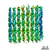
 Map data Map data | Helical reconstruction of contracted VipA/B type six secretion tubule | |||||||||
 Sample Sample |
| |||||||||
 Keywords Keywords | Type Six secretion system / tail sheath structure / Vibro cholerae / VipA/B /  gp18 / helical gp18 / helical | |||||||||
| Function / homology |  Function and homology information Function and homology information | |||||||||
| Biological species |  Vibrio cholerae O1 (bacteria) Vibrio cholerae O1 (bacteria) | |||||||||
| Method | helical reconstruction / cryo EM / Resolution: 5.8 Å | |||||||||
 Authors Authors | Kube S / Kapitein N / Zimniak T / Herzog F / Mogk A / Wendler P | |||||||||
 Citation Citation | Journal: Cell Rep / Year: 2014 Title: Structure of the VipA/B type VI secretion complex suggests a contraction-state-specific recycling mechanism. Authors: Sebastian Kube / Nicole Kapitein / Tomasz Zimniak / Franz Herzog / Axel Mogk / Petra Wendler /  Abstract: The bacterial type VI secretion system is a multicomponent molecular machine directed against eukaryotic host cells and competing bacteria. An intracellular contractile tubular structure that bears ...The bacterial type VI secretion system is a multicomponent molecular machine directed against eukaryotic host cells and competing bacteria. An intracellular contractile tubular structure that bears functional homology with bacteriophage tails is pivotal for ejection of pathogenic effectors. Here, we present the 6 Å cryoelectron microscopy structure of the contracted Vibrio cholerae tubule consisting of the proteins VipA and VipB. We localized VipA and VipB in the protomer and identified structural homology between the C-terminal segment of VipB and the tail-sheath protein of T4 phages. We propose that homologous segments in VipB and T4 phages mediate tubule contraction. We show that in type VI secretion, contraction leads to exposure of the ClpV recognition motif, which is embedded in the type VI-specific four-helix-bundle N-domain of VipB. Disaggregation of the tubules by the AAA+ protein ClpV and recycling of the VipA/B subunits are thereby limited to the contracted state. | |||||||||
| History |
|
- Structure visualization
| Movie |
Movie viewer |
|---|---|
| Structure viewer | EM map: SurfViewMolmilJmol/JSmol |
| Supplemental images |

- Downloads & links
-EMDB archive
| Map data | emd_2524.map.gz | 19 MB | EMDB map data format | |
|---|---|---|---|---|
| Header (meta data) | emd-2524-v30.xmlemd-2524.xml | 12.1 KB 12.1 KB | Display Display | EMDB header |
| Images | 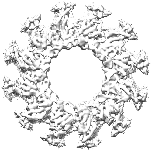 emd_2524.png emd_2524.png | 185.2 KB | ||
| Archive directory |  http://ftp.pdbj.org/pub/emdb/structures/EMD-2524ftp://ftp.pdbj.org/pub/emdb/structures/EMD-2524 http://ftp.pdbj.org/pub/emdb/structures/EMD-2524ftp://ftp.pdbj.org/pub/emdb/structures/EMD-2524 | HTTPS FTP |
-Related structure data


-Links
| EMDB pages | EMDB (EBI/PDBe) / EMDataResource |
|---|
-Map
| File | Download / File: emd_2524.map.gz / Format: CCP4 / Size: 276 MB / Type: IMAGE STORED AS FLOATING POINT NUMBER (4 BYTES) | ||||||||||||||||||||||||||||||||||||||||||||||||||||||||||||||||||||
|---|---|---|---|---|---|---|---|---|---|---|---|---|---|---|---|---|---|---|---|---|---|---|---|---|---|---|---|---|---|---|---|---|---|---|---|---|---|---|---|---|---|---|---|---|---|---|---|---|---|---|---|---|---|---|---|---|---|---|---|---|---|---|---|---|---|---|---|---|---|
| Annotation | Helical reconstruction of contracted VipA/B type six secretion tubule | ||||||||||||||||||||||||||||||||||||||||||||||||||||||||||||||||||||
| Voxel size | X=Y=Z: 1.0698 Å | ||||||||||||||||||||||||||||||||||||||||||||||||||||||||||||||||||||
| Density |
| ||||||||||||||||||||||||||||||||||||||||||||||||||||||||||||||||||||
| Symmetry | Space group: 1 | ||||||||||||||||||||||||||||||||||||||||||||||||||||||||||||||||||||
| Details | EMDB XML:
CCP4 map header:
| ||||||||||||||||||||||||||||||||||||||||||||||||||||||||||||||||||||
-Supplemental data
- Sample components
-Entire : VipA/B tubular complex in contracted state
| Entire | Name: VipA/B tubular complex in contracted state |
|---|---|
| Components |
|
-Supramolecule #1000: VipA/B tubular complex in contracted state
| Supramolecule | Name: VipA/B tubular complex in contracted state / type: sample / ID: 1000 / Details: sample forms tubular complexes of varying length / Oligomeric state: VipA and VipB form heterodimer / Number unique components: 2 |
|---|
-Macromolecule #1: VipA
| Macromolecule | Name: VipA / type: protein_or_peptide / ID: 1 / Name.synonym: TssB / Oligomeric state: heterodimer with VipB in helical array / Recombinant expression: Yes |
|---|---|
| Source (natural) | Organism: Vibrio cholerae O1 (bacteria) / Strain: ATCC 39315 / El Tor Inaba N16961 / Location in cell: cytoplasm |
| Molecular weight | Theoretical: 18.5 KDa |
| Recombinant expression | Organism: Escherichia coli K-12 (bacteria) / Recombinant strain: XL1 Blue / Recombinant plasmid: pQE31 |
| Sequence | UniProtKB: Type VI secretion system contractile sheath small subunit GO: biological_process, molecular_function, cellular_component InterPro: Type VI secretion system sheath protein TssB1 |
-Macromolecule #2: VipB
| Macromolecule | Name: VipB / type: protein_or_peptide / ID: 2 / Name.synonym: TssC / Oligomeric state: heterodimer with VipA in helical array / Recombinant expression: Yes |
|---|---|
| Source (natural) | Organism: Vibrio cholerae O1 (bacteria) / Strain: ATCC 39315 / El Tor Inaba N16961 / Location in cell: cytoplasm |
| Molecular weight | Theoretical: 55.6 KDa |
| Recombinant expression | Organism: Escherichia coli K-12 (bacteria) / Recombinant strain: XL1 Blue / Recombinant plasmid: PQE31 |
| Sequence | UniProtKB: Type VI secretion system contractile sheath large subunit GO: biological_process, molecular_function, cellular_component InterPro: Type VI secretion system TssC-like |
-Experimental details
-Structure determination
| Method | cryo EM |
|---|---|
 Processing Processing | helical reconstruction |
| Aggregation state | filament |
-Sample preparation
| Concentration | 0.3 mg/mL |
|---|---|
| Buffer | pH: 7.5 / Details: 50mM Tris, 150 mM KCl, 10 mM MgCl2 |
| Grid | Details: Quantifoil R3/3 wit carbon support film, glow discharged |
| Vitrification | Cryogen name: ETHANE / Chamber humidity: 100 % / Instrument: FEI VITROBOT MARK IV / Method: blot for 3.5 seconds before plunging |
- Electron microscopy
| Microscope | FEI TITAN KRIOS |
|---|---|
| Electron beam | Acceleration voltage: 200 kV / Electron source: FIELD EMISSION GUN |
| Electron optics | Calibrated magnification: 145821 / Illumination mode: FLOOD BEAM / Imaging mode: BRIGHT FIELDBright-field microscopy / Cs: 2.7 mm / Nominal defocus max: 4.5 µm / Nominal defocus min: 1.0 µm / Nominal magnification: 75000 |
| Sample stage | Specimen holder model: FEI TITAN KRIOS AUTOGRID HOLDER |
| Date | Jan 16, 2013 |
| Image recording | Category: CCD / Film or detector model: TVIPS TEMCAM-F416 (4k x 4k) / Digitization - Sampling interval: 15.6 µm / Number real images: 12271 / Average electron dose: 20 e/Å2 / Bits/pixel: 16 |
| Experimental equipment |  Model: Titan Krios / Image courtesy: FEI Company |
-Image processing
| CTF correction | Details: each particle |
|---|---|
| Final angle assignment | Details: theta 20 degrees, phi 60 degrees |
| Final reconstruction | Applied symmetry - Helical parameters - Δz: 22.2 Å Applied symmetry - Helical parameters - Δ&Phi: 29.44 ° Algorithm: OTHER / Resolution.type: BY AUTHOR / Resolution: 5.8 Å / Resolution method: OTHER / Software - Name: IMAGIC, SPIDER, IHRSR Details: dataset was symmetrized before final reconstruction as described in Clare & Orlova, 2012 (JSB) |
| Details | Image were processed after an adapted protocol by Clare & Orlova, 2012 (JSB) |
