Movie
Movie Controller

[English] 日本語
 Yorodumi
- EMDB-2447: Electron microscopy of the complex formed by chaperones TBCE and ... -
+ Open data

- Basic information
| Entry | Database: EMDB / ID: EMD-2447 | |||||||||
|---|---|---|---|---|---|---|---|---|---|---|
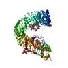
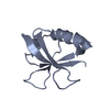
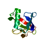
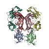
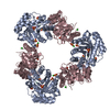
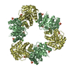
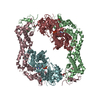
| Title | Electron microscopy of the complex formed by chaperones TBCE and TBCB and alpha-tubulin | |||||||||
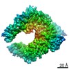
 Map data Map data | Reconstruction of the complex formed by TBCE, TBCB and alpha-tubulin | |||||||||
 Sample Sample |
| |||||||||
 Keywords Keywords | chaperones / protein folding / protein degradation / tubulin proteostasis | |||||||||
| Function / homology |  Function and homology information Function and homology informationperipheral nervous system neuron axonogenesis / post-chaperonin tubulin folding pathway / muscle atrophy / Post-chaperonin tubulin folding pathway / tubulin complex assembly / developmental growth / alpha-tubulin binding / mitotic spindle organization / adult locomotory behavior / post-embryonic development ...peripheral nervous system neuron axonogenesis / post-chaperonin tubulin folding pathway / muscle atrophy / Post-chaperonin tubulin folding pathway / tubulin complex assembly / developmental growth / alpha-tubulin binding / mitotic spindle organization / adult locomotory behavior / post-embryonic development / Hydrolases; Acting on acid anhydrides; Acting on GTP to facilitate cellular and subcellular movement / structural constituent of cytoskeleton / microtubule cytoskeleton organization / microtubule cytoskeleton / protein folding / mitotic cell cycle / nervous system development / protein-folding chaperone binding / microtubule / cell differentiation / GTPase activity / GTP binding / metal ion binding / cytoplasm / cytosol Similarity search - Function | |||||||||
| Biological species |  Homo sapiens (human) / Homo sapiens (human) /  | |||||||||
| Method | single particle reconstruction / negative staining / Resolution: 21.0 Å | |||||||||
 Authors Authors | Serna M / Carranza G / Martin-Benito J / Janowsk R / Canals A / Coll M / Zabala JC / Valpuesta JM | |||||||||
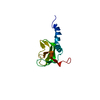
 Citation Citation | Journal: J Cell Sci / Year: 2015 Title: The structure of the complex between α-tubulin, TBCE and TBCB reveals a tubulin dimer dissociation mechanism. Authors: Marina Serna / Gerardo Carranza / Jaime Martín-Benito / Robert Janowski / Albert Canals / Miquel Coll / Juan Carlos Zabala / José María Valpuesta /  Abstract: Tubulin proteostasis is regulated by a group of molecular chaperones termed tubulin cofactors (TBC). Whereas tubulin heterodimer formation is well-characterized biochemically, its dissociation ...Tubulin proteostasis is regulated by a group of molecular chaperones termed tubulin cofactors (TBC). Whereas tubulin heterodimer formation is well-characterized biochemically, its dissociation pathway is not clearly understood. Here, we carried out biochemical assays to dissect the role of the human TBCE and TBCB chaperones in α-tubulin-β-tubulin dissociation. We used electron microscopy and image processing to determine the three-dimensional structure of the human TBCE, TBCB and α-tubulin (αEB) complex, which is formed upon α-tubulin-β-tubulin heterodimer dissociation by the two chaperones. Docking the atomic structures of domains of these proteins, including the TBCE UBL domain, as we determined by X-ray crystallography, allowed description of the molecular architecture of the αEB complex. We found that heterodimer dissociation is an energy-independent process that takes place through a disruption of the α-tubulin-β-tubulin interface that is caused by a steric interaction between β-tubulin and the TBCE cytoskeleton-associated protein glycine-rich (CAP-Gly) and leucine-rich repeat (LRR) domains. The protruding arrangement of chaperone ubiquitin-like (UBL) domains in the αEB complex suggests that there is a direct interaction of this complex with the proteasome, thus mediating α-tubulin degradation. | |||||||||
| History |
|
- Structure visualization
| Movie |
Movie viewer |
|---|---|
| Structure viewer | EM map: SurfViewMolmilJmol/JSmol |
| Supplemental images |

- Downloads & links
-EMDB archive
| Map data | emd_2447.map.gz | 119.2 KB | EMDB map data format | |
|---|---|---|---|---|
| Header (meta data) | emd-2447-v30.xmlemd-2447.xml | 14.9 KB 14.9 KB | Display Display | EMDB header |
| Images | 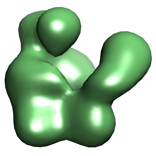 EMD-2447.png EMD-2447.png | 91.2 KB | ||
| Archive directory |  http://ftp.pdbj.org/pub/emdb/structures/EMD-2447ftp://ftp.pdbj.org/pub/emdb/structures/EMD-2447 http://ftp.pdbj.org/pub/emdb/structures/EMD-2447ftp://ftp.pdbj.org/pub/emdb/structures/EMD-2447 | HTTPS FTP |
-Validation report
| Summary document | emd_2447_validation.pdf.gz | 213.6 KB | Display | EMDB validaton report |
|---|---|---|---|---|
| Full document | emd_2447_full_validation.pdf.gz | 212.7 KB | Display | |
| Data in XML | emd_2447_validation.xml.gz | 4.5 KB | Display | |
| Arichive directory | https://ftp.pdbj.org/pub/emdb/validation_reports/EMD-2447ftp://ftp.pdbj.org/pub/emdb/validation_reports/EMD-2447 | HTTPS FTP |
-Related structure data


-Links
| EMDB pages | EMDB (EBI/PDBe) / EMDataResource |
|---|---|
| Related items in Molecule of the Month |


-Map
| File | Download / File: emd_2447.map.gz / Format: CCP4 / Size: 126 KB / Type: IMAGE STORED AS FLOATING POINT NUMBER (4 BYTES) | ||||||||||||||||||||||||||||||||||||||||||||||||||||||||||||
|---|---|---|---|---|---|---|---|---|---|---|---|---|---|---|---|---|---|---|---|---|---|---|---|---|---|---|---|---|---|---|---|---|---|---|---|---|---|---|---|---|---|---|---|---|---|---|---|---|---|---|---|---|---|---|---|---|---|---|---|---|---|
| Annotation | Reconstruction of the complex formed by TBCE, TBCB and alpha-tubulin | ||||||||||||||||||||||||||||||||||||||||||||||||||||||||||||
| Voxel size | X=Y=Z: 4.66 Å | ||||||||||||||||||||||||||||||||||||||||||||||||||||||||||||
| Density |
| ||||||||||||||||||||||||||||||||||||||||||||||||||||||||||||
| Symmetry | Space group: 1 | ||||||||||||||||||||||||||||||||||||||||||||||||||||||||||||
| Details | EMDB XML:
CCP4 map header:
| ||||||||||||||||||||||||||||||||||||||||||||||||||||||||||||
-Supplemental data
- Sample components
-Entire : Ternary complex of alpha-tubulin, TBCE and TBCB
| Entire | Name: Ternary complex of alpha-tubulin, TBCE and TBCB |
|---|---|
| Components |
|
-Supramolecule #1000: Ternary complex of alpha-tubulin, TBCE and TBCB
| Supramolecule | Name: Ternary complex of alpha-tubulin, TBCE and TBCB / type: sample / ID: 1000 / Oligomeric state: heterotrimer / Number unique components: 3 |
|---|---|
| Molecular weight | Theoretical: 137 KDa |
-Macromolecule #1: Tubulin binding cofactor E
| Macromolecule | Name: Tubulin binding cofactor E / type: protein_or_peptide / ID: 1 / Name.synonym: Tubulin-specific chaperone E / Number of copies: 1 / Oligomeric state: Monomer / Recombinant expression: Yes |
|---|---|
| Source (natural) | Organism: Homo sapiens (human) / synonym: Human / Location in cell: Cytoplasm |
| Molecular weight | Theoretical: 59 KDa |
| Recombinant expression | Organism:   Spodoptera frugiperda (fall armyworm) / Recombinant cell: Sf9 / Recombinant plasmid: pFastBac-1 Spodoptera frugiperda (fall armyworm) / Recombinant cell: Sf9 / Recombinant plasmid: pFastBac-1 |
| Sequence | UniProtKB: Tubulin-specific chaperone E / InterPro: CAP Gly-rich domain |
-Macromolecule #2: Tubulin binding cofactor B
| Macromolecule | Name: Tubulin binding cofactor B / type: protein_or_peptide / ID: 2 Name.synonym: Tubulin-specific chaperone B, Tubulin-folding cofactor B Number of copies: 1 / Oligomeric state: Monomer / Recombinant expression: Yes |
|---|---|
| Source (natural) | Organism: Homo sapiens (human) / synonym: Human / Location in cell: Cytoplasm |
| Molecular weight | Theoretical: 27 KDa |
| Recombinant expression | Organism:  |
| Sequence | UniProtKB: Tubulin-folding cofactor B / InterPro: CAP Gly-rich domain |
-Macromolecule #3: alpha-tubulin
| Macromolecule | Name: alpha-tubulin / type: protein_or_peptide / ID: 3 / Name.synonym: Tubulin alpha-1B chain / Number of copies: 1 / Oligomeric state: Monomer / Recombinant expression: No |
|---|---|
| Source (natural) | Organism: |
| Molecular weight | Theoretical: 50 KDa |
| Sequence | UniProtKB: Tubulin alpha-1B chain / InterPro: Alpha tubulin |
-Experimental details
-Structure determination
| Method | negative staining |
|---|---|
 Processing Processing | single particle reconstruction |
| Aggregation state | particle |
-Sample preparation
| Concentration | 0.02 mg/mL |
|---|---|
| Buffer | pH: 6.7 / Details: 100mM MES-NaOH, 25mM KCl, 1mM MgCl2, 1mM EGTA |
| Staining | Type: NEGATIVE Details: Grids with adsorbed protein floated on 2% w/v uranyl acetate for 1 min. |
| Grid | Details: 300 mesh copper grid with thin carbon support, glow discharged in air atmosphere |
| Vitrification | Cryogen name: NONE / Instrument: OTHER |
- Electron microscopy
| Microscope | JEOL 1200EXII |
|---|---|
| Alignment procedure | Legacy - Astigmatism: Objective lens astigmatism was corrected at 100,000 times magnification |
| Date | Nov 1, 2010 |
| Image recording | Category: FILM / Film or detector model: KODAK SO-163 FILM / Digitization - Scanner: ZEISS SCAI / Digitization - Sampling interval: 14 µm / Number real images: 85 / Average electron dose: 10 e/Å2 |
| Electron beam | Acceleration voltage: 100 kV / Electron source: TUNGSTEN HAIRPIN |
| Electron optics | Illumination mode: FLOOD BEAM / Imaging mode: BRIGHT FIELD / Cs: 5.6 mm / Nominal defocus max: 1.5 µm / Nominal defocus min: 1.0 µm / Nominal magnification: 60000 |
| Sample stage | Specimen holder model: JEOL |
-Image processing
| Details | The particles were manually selected using the XMIPP program |
|---|---|
| CTF correction | Details: N. Grigorieff CTFFIND3 |
| Final reconstruction | Applied symmetry - Point group: C1 (asymmetric) / Algorithm: OTHER / Resolution.type: BY AUTHOR / Resolution: 21.0 Å / Resolution method: FSC 0.33 CUT-OFF / Software - Name: EMAN, Spider, XMIPP Details: Individual particles were manually selected using XMIPP software package. Number images used: 26129 |
-Atomic model buiding 1
| Initial model | PDB ID: Chain - Chain ID: A |
|---|---|
| Software | Name:  Chimera Chimera |
| Details | The domain was fitted by manual docking using Chimera |
| Refinement | Space: REAL / Protocol: RIGID BODY FIT |

-Atomic model buiding 2
| Initial model | PDB ID: Chain - Chain ID: A |
|---|---|
| Software | Name: Chimera |
| Details | The domain was fitted by manual docking using Chimera |
| Refinement | Space: REAL / Protocol: RIGID BODY FIT |
-Atomic model buiding 3
| Initial model | PDB ID: Chain - Chain ID: A |
|---|---|
| Software | Name: Chimera |
| Details | The domain was fitted by manual docking using Chimera |
| Refinement | Space: REAL / Protocol: RIGID BODY FIT |

-Atomic model buiding 4
| Initial model | PDB ID: Chain - Chain ID: A |
|---|---|
| Software | Name: Chimera |
| Details | The domain was fitted by manual docking using Chimera |
| Refinement | Space: REAL / Protocol: RIGID BODY FIT |
-Atomic model buiding 5
| Initial model | PDB ID: Chain - Chain ID: A |
|---|---|
| Software | Name: Chimera |
| Details | The domain was fitted by manual docking using Chimera |
| Refinement | Space: REAL / Protocol: RIGID BODY FIT |