 Movie
Movie Controller


+ Open data

- Basic information
| Entry | Database: EMDB / ID: EMD-2412 | |||||||||
|---|---|---|---|---|---|---|---|---|---|---|
| Title | cryo-electron tomography of poliovirus-PVR-liposome complex | |||||||||
 Map data Map data | Subtomogram asymmetric average of all 651 virus particles attached to membrane. | |||||||||
 Sample Sample |
| |||||||||
 Keywords Keywords | poliovirus PVR poliovirus receptor liposome 135S | |||||||||
| Biological species |    Human poliovirus 1 Mahoney Human poliovirus 1 Mahoney | |||||||||
| Method | subtomogram averaging / cryo EM | |||||||||
 Authors Authors | Strauss M / Levy HC / Bostina M / Filman DJ / Hogle JM | |||||||||
 Citation Citation | Journal: J Virol / Year: 2013 Title: RNA transfer from poliovirus 135S particles across membranes is mediated by long umbilical connectors. Authors: Mike Strauss / Hazel C Levy / Mihnea Bostina / David J Filman / James M Hogle /  Abstract: During infection, the binding of poliovirus to its cell surface receptor at 37°C triggers an expansion of the virus in which internal polypeptides that bind to membranes are externalized. ...During infection, the binding of poliovirus to its cell surface receptor at 37°C triggers an expansion of the virus in which internal polypeptides that bind to membranes are externalized. Subsequently, in a poorly understood process, the viral RNA genome is transferred directly across an endosomal membrane, and into the host cell cytoplasm, to initiate infection. Here, cryoelectron tomography demonstrates the results of 37°C warming of a poliovirus-receptor-liposome model complex that was produced using Ni-nitrilotriacetic acid lipids and His-tagged receptor ectodomains. In total, 651 subtomographic volumes were aligned, classified, and averaged to obtain detailed pictures, showing both the conversion of virus into its expanded form and the passage of RNA into intact liposomes. Unexpectedly, the virus and membrane surfaces were located ∼50 Å apart, with the 5-fold axis tilted away from the perpendicular, and the solvent spaces between them were spanned by either one or two long "umbilical" density features that lie at an angle to the virus and membrane. The thinner connector, which sometimes appears alone, is 28 to 30 Å in diameter and has a footprint on the virus surface located close to either a 5-fold or a 3-fold axis. The broader connector has a footprint near the quasi-3-fold hole that opens upon virus expansion and is hypothesized to include RNA, shielded from enzymatic degradation by polypeptides that include the N-terminal extension of VP1 and capsid protein VP4. The implications of these observations for the mechanism of RNase-protected RNA transfer in picornaviruses are discussed. | |||||||||
| History |
|
- Structure visualization
| Movie |
 Movie viewer Movie viewer |
|---|---|
| Structure viewer | EM map: SurfViewMolmilJmol/JSmol |
| Supplemental images |

- Downloads & links
-EMDB archive
| Map data | emd_2412.map.gz | 7.1 MB | EMDB map data format | |
|---|---|---|---|---|
| Header (meta data) | emd-2412-v30.xmlemd-2412.xml | 9 KB 9 KB | Display Display | EMDB header |
| Images | 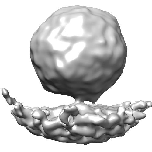 emd_2412.jpg emd_2412.jpg | 21.4 KB | ||
| Archive directory |  http://ftp.pdbj.org/pub/emdb/structures/EMD-2412ftp://ftp.pdbj.org/pub/emdb/structures/EMD-2412 http://ftp.pdbj.org/pub/emdb/structures/EMD-2412ftp://ftp.pdbj.org/pub/emdb/structures/EMD-2412 | HTTPS FTP |
-Related structure data

-Links
| EMDB pages | EMDB (EBI/PDBe) / EMDataResource |
|---|---|
| Related items in Molecule of the Month |

-Map
| File | Download / File: emd_2412.map.gz / Format: CCP4 / Size: 7.8 MB / Type: IMAGE STORED AS FLOATING POINT NUMBER (4 BYTES) | ||||||||||||||||||||||||||||||||||||||||||||||||||||||||||||||||||||
|---|---|---|---|---|---|---|---|---|---|---|---|---|---|---|---|---|---|---|---|---|---|---|---|---|---|---|---|---|---|---|---|---|---|---|---|---|---|---|---|---|---|---|---|---|---|---|---|---|---|---|---|---|---|---|---|---|---|---|---|---|---|---|---|---|---|---|---|---|---|
| Annotation | Subtomogram asymmetric average of all 651 virus particles attached to membrane. | ||||||||||||||||||||||||||||||||||||||||||||||||||||||||||||||||||||
| Voxel size | X=Y=Z: 6.25 Å | ||||||||||||||||||||||||||||||||||||||||||||||||||||||||||||||||||||
| Density |
| ||||||||||||||||||||||||||||||||||||||||||||||||||||||||||||||||||||
| Symmetry | Space group: 1 | ||||||||||||||||||||||||||||||||||||||||||||||||||||||||||||||||||||
| Details | EMDB XML:
CCP4 map header:
| ||||||||||||||||||||||||||||||||||||||||||||||||||||||||||||||||||||
-Supplemental data
- Sample components
-Entire : 135S poliovirus - membrane complex
| Entire | Name: 135S poliovirus - membrane complex |
|---|---|
| Components |
|
-Supramolecule #1000: 135S poliovirus - membrane complex
| Supramolecule | Name: 135S poliovirus - membrane complex / type: sample / ID: 1000 Oligomeric state: one particle connects to one membrane at 2 sites Number unique components: 1 |
|---|
-Supramolecule #1: Human poliovirus 1 Mahoney
| Supramolecule | Name: Human poliovirus 1 Mahoney / type: virus / ID: 1 Details: The particle is in the expanded (135S) state, and connected to the membrane. NCBI-ID: 12081 / Sci species name: Human poliovirus 1 Mahoney / Virus type: VIRION / Virus isolate: SEROTYPE / Virus enveloped: No / Virus empty: No / Sci species serotype: PV-1 |
|---|---|
| Host (natural) | Organism:  Homo sapiens (human) / synonym: VERTEBRATES Homo sapiens (human) / synonym: VERTEBRATES |
| Molecular weight | Experimental: 8.5 MDa / Theoretical: 8.5 MDa |
| Virus shell | Shell ID: 1 / Diameter: 330 Å / T number (triangulation number): 1 |
-Experimental details
-Structure determination
| Method | cryo EM |
|---|---|
 Processing Processing | subtomogram averaging |
| Aggregation state | particle |
-Sample preparation
| Concentration | 1 mg/mL |
|---|---|
| Buffer | pH: 7.3 / Details: 50mM Hepes, 50mM NaCl |
| Grid | Details: 200 mesh copper Quantifoil grids (R2/2) with 3 nm carbon support on top. |
| Vitrification | Cryogen name: ETHANE / Chamber humidity: 90 % / Chamber temperature: 120 K / Instrument: FEI VITROBOT MARK III / Method: 2 second blot |
- Electron microscopy
| Microscope | FEI TITAN KRIOS |
|---|---|
| Electron beam | Acceleration voltage: 300 kV / Electron source: FIELD EMISSION GUN |
| Electron optics | Calibrated magnification: 45454 / Illumination mode: FLOOD BEAM / Imaging mode: BRIGHT FIELDBright-field microscopy / Cs: 2.7 mm / Nominal magnification: 50000 |
| Sample stage | Specimen holder model: FEI TITAN KRIOS AUTOGRID HOLDER |
| Alignment procedure | Legacy - Electron beam tilt params: 0 |
| Date | Oct 10, 2010 |
| Image recording | Category: CCD / Film or detector model: GATAN ULTRASCAN 1000 (2k x 2k) / Average electron dose: 60 e/Å2 / Bits/pixel: 16 |
| Experimental equipment |  Model: Titan Krios / Image courtesy: FEI Company |
-Image processing
| Final reconstruction | Applied symmetry - Point group: C1 (asymmetric) / Software - Name: IMOD, PEET, BSOFT, EMAN2, SPARX / Number subtomograms used: 651 |
|---|---|
| Details | all particles close to a membrane were averaged |
