Movie
Movie Controller


+ Open data

- Basic information
| Entry | Database: EMDB / ID: EMD-2365 | |||||||||
|---|---|---|---|---|---|---|---|---|---|---|
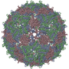
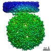
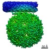
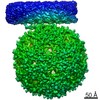
| Title | Asymmetric structure of a virus-receptor complex | |||||||||
 Map data Map data | Sub-tomogram average of bacteriophage MS2 bound to its receptor | |||||||||
 Sample Sample |
| |||||||||
 Keywords Keywords |  virus / receptor / complex / asymmetric / bacteriophage virus / receptor / complex / asymmetric / bacteriophage | |||||||||
| Function / homology |  Function and homology information Function and homology informationnegative regulation of viral translation / T=3 icosahedral viral capsid / regulation of translation / structural molecule activity / RNA binding / identical protein bindingSimilarity search - Function | |||||||||
| Biological species |  Escherichia coli (E. coli) / Escherichia coli (E. coli) /  Enterobacterio phage MS2 (virus) Enterobacterio phage MS2 (virus) | |||||||||
| Method | subtomogram averaging / cryo EM / Resolution: 39.0 Å | |||||||||
 Authors Authors | Dent KC / Thompson R / Barker AM / Barr JN / Hiscox JA / Stockley PG / Ranson NA | |||||||||
 Citation Citation | Journal: Structure / Year: 2013 Title: The asymmetric structure of an icosahedral virus bound to its receptor suggests a mechanism for genome release. Authors: Kyle C Dent / Rebecca Thompson / Amy M Barker / Julian A Hiscox / John N Barr / Peter G Stockley / Neil A Ranson /  Abstract: Simple, spherical RNA viruses have well-understood, symmetric protein capsids, but little structural information is available for their asymmetric components, such as minor proteins and their ...Simple, spherical RNA viruses have well-understood, symmetric protein capsids, but little structural information is available for their asymmetric components, such as minor proteins and their genomes, which are vital for infection. Here, we report an asymmetric structure of bacteriophage MS2, attached to its receptor, the F-pilus. Cryo-electron tomography and subtomographic averaging of such complexes result in a structure containing clear density for the packaged genome, implying that the conformation of the genome is the same in each virus particle. The data also suggest that the single-copy viral maturation protein breaks the symmetry of the capsid, occupying a position that would be filled by a coat protein dimer in an icosahedral shell. This capsomere can thus fulfill its known biological roles in receptor and genome binding and suggests an exit route for the genome during infection. | |||||||||
| History |
|
- Structure visualization
| Movie |
Movie viewer |
|---|---|
| Structure viewer | EM map: SurfViewMolmilJmol/JSmol |
| Supplemental images |

- Downloads & links
-EMDB archive
| Map data | emd_2365.map.gz | 895.9 KB | EMDB map data format | |
|---|---|---|---|---|
| Header (meta data) | emd-2365-v30.xmlemd-2365.xml | 9.5 KB 9.5 KB | Display Display | EMDB header |
| Images | 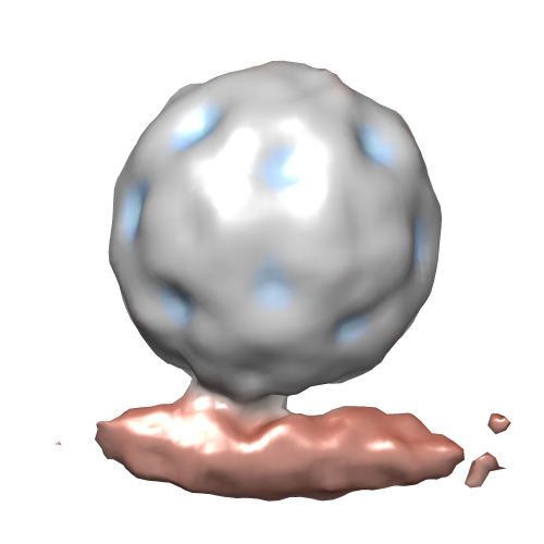 emd_2365.png emd_2365.png | 78.6 KB | ||
| Archive directory |  http://ftp.pdbj.org/pub/emdb/structures/EMD-2365ftp://ftp.pdbj.org/pub/emdb/structures/EMD-2365 http://ftp.pdbj.org/pub/emdb/structures/EMD-2365ftp://ftp.pdbj.org/pub/emdb/structures/EMD-2365 | HTTPS FTP |
-Related structure data
| Related structure data |  4bp7MC M: atomic model generated by this map C: citing same article ( |
|---|---|
| Similar structure data |


-Links
| EMDB pages | EMDB (EBI/PDBe) / EMDataResource |
|---|---|
| Related items in Molecule of the Month |

-Map
| File | Download / File: emd_2365.map.gz / Format: CCP4 / Size: 1001 KB / Type: IMAGE STORED AS FLOATING POINT NUMBER (4 BYTES) | ||||||||||||||||||||||||||||||||||||||||||||||||||||||||||||||||||||
|---|---|---|---|---|---|---|---|---|---|---|---|---|---|---|---|---|---|---|---|---|---|---|---|---|---|---|---|---|---|---|---|---|---|---|---|---|---|---|---|---|---|---|---|---|---|---|---|---|---|---|---|---|---|---|---|---|---|---|---|---|---|---|---|---|---|---|---|---|---|
| Annotation | Sub-tomogram average of bacteriophage MS2 bound to its receptor | ||||||||||||||||||||||||||||||||||||||||||||||||||||||||||||||||||||
| Voxel size | X=Y=Z: 9.12 Å | ||||||||||||||||||||||||||||||||||||||||||||||||||||||||||||||||||||
| Density |
| ||||||||||||||||||||||||||||||||||||||||||||||||||||||||||||||||||||
| Symmetry | Space group: 1 | ||||||||||||||||||||||||||||||||||||||||||||||||||||||||||||||||||||
| Details | EMDB XML:
CCP4 map header:
| ||||||||||||||||||||||||||||||||||||||||||||||||||||||||||||||||||||
-Supplemental data
- Sample components
-Entire : Bacteriophage MS2 bound to its receptor, the E. coli F-pilus
| Entire | Name: Bacteriophage MS2 bound to its receptor, the E. coli F-pilus |
|---|---|
| Components |
|
-Supramolecule #1000: Bacteriophage MS2 bound to its receptor, the E. coli F-pilus
| Supramolecule | Name: Bacteriophage MS2 bound to its receptor, the E. coli F-pilus type: sample / ID: 1000 / Details: Sub-tomographic averaging of virus-decorated pili Oligomeric state: One T=3 icosahedral virus bound to a pilus fibre Number unique components: 2 |
|---|
-Supramolecule #1: Enterobacterio phage MS2
| Supramolecule | Name: Enterobacterio phage MS2 / type: virus / ID: 1 / NCBI-ID: 12022 / Sci species name: Enterobacterio phage MS2 / Virus type: VIRION / Virus isolate: SPECIES / Virus enveloped: No / Virus empty: No |
|---|---|
| Host (natural) | Organism: Escherichia coli (E. coli) / synonym: BACTERIA(EUBACTERIA) |
| Virus shell | Shell ID: 1 / Diameter: 290 Å / T number (triangulation number): 3 |
-Supramolecule #2: F-Pilus
| Supramolecule | Name: F-Pilus / type: organelle_or_cellular_component / ID: 2 / Recombinant expression: No |
|---|---|
| Source (natural) | Organism: Escherichia coli (E. coli) |
-Experimental details
-Structure determination
| Method | cryo EM |
|---|---|
 Processing Processing | subtomogram averaging |
| Aggregation state | particle |
-Sample preparation
| Grid | Details: 200 mesh lacey carbon grids, glow discharged in air. |
|---|---|
| Vitrification | Cryogen name: ETHANE / Instrument: HOMEMADE PLUNGER |
- Electron microscopy
| Microscope | FEI TECNAI 12 |
|---|---|
| Electron beam | Acceleration voltage: 120 kV / Electron source: LAB6 |
| Electron optics | Calibrated magnification: 23000 / Illumination mode: FLOOD BEAM / Imaging mode: BRIGHT FIELDBright-field microscopy / Nominal defocus max: 3.0 µm / Nominal defocus min: 3.0 µm / Nominal magnification: 23000 |
| Sample stage | Specimen holder model: GATAN LIQUID NITROGEN / Tilt series - Axis1 - Min angle: -60 ° / Tilt series - Axis1 - Max angle: 60 ° |
| Date | Jan 1, 2012 |
| Image recording | Category: CCD / Film or detector model: GATAN ULTRASCAN 1000 (2k x 2k) |
-Image processing
| Final reconstruction | Applied symmetry - Point group: C1 (asymmetric) / Resolution.type: BY AUTHOR / Resolution: 39.0 Å / Resolution method: FSC 0.5 CUT-OFF / Software - Name: IMOD, PEET / Number subtomograms used: 1000 |
|---|