 Movie
Movie Controller

[English] 日本語
 Yorodumi
- EMDB-2224: Negative stain microscopy of a dimer of Actin-related protein 8 (... -
+ Open data

- Basic information
| Entry | Database: EMDB / ID: EMD-2224 | |||||||||
|---|---|---|---|---|---|---|---|---|---|---|
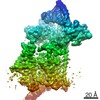
| Title | Negative stain microscopy of a dimer of Actin-related protein 8 (Arp8) from S. cerevisiae. | |||||||||
 Map data Map data | Negative stain reconstruction of a dimeric form of full-length Actin Related Protein 8 (Arp8) from Saccharomyces cerevisiae. | |||||||||
 Sample Sample |
| |||||||||
 Keywords Keywords |  Chromatin remodelling / INO80 complex / Actin related protein / Arp8 Chromatin remodelling / INO80 complex / Actin related protein / Arp8 | |||||||||
| Biological species |  Saccharomyces cerevisiae (brewer's yeast) Saccharomyces cerevisiae (brewer's yeast) | |||||||||
| Method | single particle reconstruction / negative staining / Resolution: 22.0 Å | |||||||||
 Authors Authors | Bose DA / Saravanan M / Wuerges J / McCormack EA / Cook NJ / Zhang X / Wigley DB | |||||||||
 Citation Citation | Journal: Proc Natl Acad Sci U S A / Year: 2012 Title: Interactions between the nucleosome histone core and Arp8 in the INO80 chromatin remodeling complex. Authors: Matheshwaran Saravanan / Jochen Wuerges / Daniel Bose / Elizabeth A McCormack / Nicola J Cook / Xiaodong Zhang / Dale B Wigley /  Abstract: Actin-related protein Arp8 is a component of the INO80 chromatin remodeling complex. Yeast Arp8 (yArp8) comprises two domains: a 25-KDa N-terminal domain, found only in yeast, and a 75-KDa C-terminal ...Actin-related protein Arp8 is a component of the INO80 chromatin remodeling complex. Yeast Arp8 (yArp8) comprises two domains: a 25-KDa N-terminal domain, found only in yeast, and a 75-KDa C-terminal domain (yArp8CTD) that contains the actin fold and is conserved across other species. The crystal structure shows that yArp8CTD contains three insertions within the actin core. Using a combination of biochemistry and EM, we show that Arp8 forms a complex with nucleosomes, and that the principal interactions are via the H3 and H4 histones, mediated through one of the yArp8 insertions. We show that recombinant yArp8 exists in monomeric and dimeric states, but the dimer is the biologically relevant form required for stable interactions with histones that exploits the twofold symmetry of the nucleosome core. Taken together, these data provide unique insight into the stoichiometry, architecture, and molecular interactions between components of the INO80 remodeling complex and nucleosomes, providing a first step toward building up the structure of the complex. | |||||||||
| History |
|
- Structure visualization
| Movie |
 Movie viewer Movie viewer |
|---|---|
| Structure viewer | EM map: SurfViewMolmilJmol/JSmol |
| Supplemental images |

- Downloads & links
-EMDB archive
| Map data | emd_2224.map.gz | 414.9 KB | EMDB map data format | |
|---|---|---|---|---|
| Header (meta data) | emd-2224-v30.xmlemd-2224.xml | 9.9 KB 9.9 KB | Display Display | EMDB header |
| Images | emd_2224.tif | 656.9 KB | ||
| Archive directory |  http://ftp.pdbj.org/pub/emdb/structures/EMD-2224ftp://ftp.pdbj.org/pub/emdb/structures/EMD-2224 http://ftp.pdbj.org/pub/emdb/structures/EMD-2224ftp://ftp.pdbj.org/pub/emdb/structures/EMD-2224 | HTTPS FTP |
-Related structure data


-Links
| EMDB pages | EMDB (EBI/PDBe) / EMDataResource |
|---|
-Map
| File | Download / File: emd_2224.map.gz / Format: CCP4 / Size: 7.8 MB / Type: IMAGE STORED AS FLOATING POINT NUMBER (4 BYTES) | ||||||||||||||||||||||||||||||||||||||||||||||||||||||||||||||||||||
|---|---|---|---|---|---|---|---|---|---|---|---|---|---|---|---|---|---|---|---|---|---|---|---|---|---|---|---|---|---|---|---|---|---|---|---|---|---|---|---|---|---|---|---|---|---|---|---|---|---|---|---|---|---|---|---|---|---|---|---|---|---|---|---|---|---|---|---|---|---|
| Annotation | Negative stain reconstruction of a dimeric form of full-length Actin Related Protein 8 (Arp8) from Saccharomyces cerevisiae. | ||||||||||||||||||||||||||||||||||||||||||||||||||||||||||||||||||||
| Voxel size | X=Y=Z: 3.52 Å | ||||||||||||||||||||||||||||||||||||||||||||||||||||||||||||||||||||
| Density |
| ||||||||||||||||||||||||||||||||||||||||||||||||||||||||||||||||||||
| Symmetry | Space group: 1 | ||||||||||||||||||||||||||||||||||||||||||||||||||||||||||||||||||||
| Details | EMDB XML:
CCP4 map header:
| ||||||||||||||||||||||||||||||||||||||||||||||||||||||||||||||||||||
-Supplemental data
- Sample components
-Entire : Dimeric form of full-length Actin Related Protein 8 (Arp8) from S...
| Entire | Name: Dimeric form of full-length Actin Related Protein 8 (Arp8) from Saccharomyces cerevisiae. |
|---|---|
| Components |
|
-Supramolecule #1000: Dimeric form of full-length Actin Related Protein 8 (Arp8) from S...
| Supramolecule | Name: Dimeric form of full-length Actin Related Protein 8 (Arp8) from Saccharomyces cerevisiae. type: sample / ID: 1000 / Oligomeric state: dimer / Number unique components: 1 |
|---|---|
| Molecular weight | Experimental: 217 KDa / Theoretical: 200 KDa Method: Multi angle light scattering (MALS) (0.231MDa) and Analytical ultracentrifugation (AUC) (0.217MDa) |
-Macromolecule #1: Actin-like protein ARP8
| Macromolecule | Name: Actin-like protein ARP8 / type: protein_or_peptide / ID: 1 / Number of copies: 2 / Oligomeric state: dimer / Recombinant expression: Yes |
|---|---|
| Source (natural) | Organism: Saccharomyces cerevisiae (brewer's yeast) / synonym: Baker's yeast / Location in cell: Nucleus |
| Molecular weight | Theoretical: 100 KDa |
| Recombinant expression | Organism:  Escherichia coli (E. coli) Escherichia coli (E. coli) |
-Experimental details
-Structure determination
| Method | negative staining |
|---|---|
 Processing Processing | single particle reconstruction |
| Aggregation state | particle |
-Sample preparation
| Concentration | 0.05 mg/mL |
|---|---|
| Staining | Type: NEGATIVE Details: 2 ul of ~0.05mg/ml Full length Arp8 was applied to glow-discharged continuous carbon grids (TAAB). Sample was adsorbed for 20s, then stained with 2% w/v uranyl acetate for 40s before blotting and air drying. |
| Grid | Details: Copper 300 mesh continuous carbon grids (TAAB) |
| Vitrification | Cryogen name: NONE / Instrument: OTHER |
- Electron microscopy
| Microscope | FEI/PHILIPS CM200FEG |
|---|---|
| Electron beam | Acceleration voltage: 200 kV / Electron source: FIELD EMISSION GUN |
| Electron optics | Illumination mode: SPOT SCAN / Imaging mode: BRIGHT FIELDBright-field microscopy / Cs: 2.1 mm / Nominal defocus max: 2.5 µm / Nominal defocus min: 1.0 µm / Nominal magnification: 50000 |
| Sample stage | Specimen holder: single tilt / Specimen holder model: SIDE ENTRY, EUCENTRIC |
| Date | Mar 23, 2010 |
| Image recording | Category: CCD / Film or detector model: TVIPS TEMCAM-F415 (4k x 4k) / Average electron dose: 10 e/Å2 Details: Data collected on a 4k x 4k CCD camera at 50000x magnification. Sampling interval was 1.76A/pixel. Bits/pixel: 16 |
-Image processing
| CTF correction | Details: Each particle |
|---|---|
| Final angle assignment | Details: Imagic |
| Final reconstruction | Applied symmetry - Point group: C2 (2 fold cyclic) / Algorithm: OTHER / Resolution.type: BY AUTHOR / Resolution: 22.0 Å / Resolution method: FSC 0.5 CUT-OFF / Software - Name: IMAGIC, V, TIGRIS, EMAN / Number images used: 3680 |
-Atomic model buiding 1
| Initial model | PDB ID: |
|---|---|
| Software | Name: Chimera, CNS, Situs |
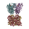
| Details | Protocol: Rigid body. The Arp8 crystal structure was positioned interactively in Chimera, then the fit was refined using Situs and CNS. |
| Refinement | Space: REAL / Protocol: RIGID BODY FIT |
