Movie
Movie Controller

[English] 日本語
 Yorodumi
- EMDB-2205: Cryo-electron microscopy reconstruction of the helical part of in... -
+ Open data

- Basic information
| Entry | Database: EMDB / ID: EMD-2205 | |||||||||
|---|---|---|---|---|---|---|---|---|---|---|
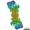
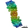
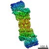
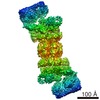
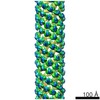
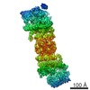
| Title | Cryo-electron microscopy reconstruction of the helical part of influenza A virus ribonucleoprotein isolated from virions. | |||||||||
 Map data Map data | Reconstruction of the helical part of the influeza A virus ribonucleoprotein | |||||||||
 Sample Sample |
| |||||||||
 Keywords Keywords |  Influenza / ribonucleoprotein / nucleoprotein / nucleocapsid / RNA / helical structure Influenza / ribonucleoprotein / nucleoprotein / nucleocapsid / RNA / helical structure | |||||||||
| Function / homology |  Function and homology information Function and homology informationnegative stranded viral RNA replication / helical viral capsid / viral penetration into host nucleus / viral nucleocapsid / symbiont entry into host cell / ribonucleoprotein complex / host cell nucleus / structural molecule activity / RNA binding / identical protein bindingSimilarity search - Function | |||||||||
| Biological species |   Influenza A virus Influenza A virus | |||||||||
| Method | helical reconstruction / cryo EM / negative staining / Resolution: 18.0 Å | |||||||||
 Authors Authors | Arranz R / Coloma R / Chichon FJ / Conesa JJ / Carrascosa JL / Valpuesta JM / Ortin J / Martin-Benito J | |||||||||
 Citation Citation | Journal: Science / Year: 2012 Title: The structure of native influenza virion ribonucleoproteins. Authors: Rocío Arranz / Rocío Coloma / Francisco Javier Chichón / José Javier Conesa / José L Carrascosa / José M Valpuesta / Juan Ortín / Jaime Martín-Benito /  Abstract: The influenza viruses cause annual epidemics of respiratory disease and occasional pandemics, which constitute a major public-health issue. The segmented negative-stranded RNAs are associated with ...The influenza viruses cause annual epidemics of respiratory disease and occasional pandemics, which constitute a major public-health issue. The segmented negative-stranded RNAs are associated with the polymerase complex and nucleoprotein (NP), forming ribonucleoproteins (RNPs), which are responsible for virus transcription and replication. We describe the structure of native RNPs derived from virions. They show a double-helical conformation in which two NP strands of opposite polarity are associated with each other along the helix. Both strands are connected by a short loop at one end of the particle and interact with the polymerase complex at the other end. This structure will be relevant for unraveling the mechanisms of nuclear import of parental virus RNPs, their transcription and replication, and the encapsidation of progeny RNPs into virions. | |||||||||
| History |
|
- Structure visualization
| Movie |
Movie viewer |
|---|---|
| Structure viewer | EM map: SurfViewMolmilJmol/JSmol |
| Supplemental images |

- Downloads & links
-EMDB archive
| Map data | emd_2205.map.gz | 578.5 KB | EMDB map data format | |
|---|---|---|---|---|
| Header (meta data) | emd-2205-v30.xmlemd-2205.xml | 10.7 KB 10.7 KB | Display Display | EMDB header |
| Images | EMD-2205.tif | 238.8 KB | ||
| Archive directory |  http://ftp.pdbj.org/pub/emdb/structures/EMD-2205ftp://ftp.pdbj.org/pub/emdb/structures/EMD-2205 http://ftp.pdbj.org/pub/emdb/structures/EMD-2205ftp://ftp.pdbj.org/pub/emdb/structures/EMD-2205 | HTTPS FTP |
-Related structure data
| Related structure data |  4bblMC  2206C  2207C  2208C M: atomic model generated by this map C: citing same article ( |
|---|---|
| Similar structure data |


-Links
| EMDB pages | EMDB (EBI/PDBe) / EMDataResource |
|---|
-Map
| File | Download / File: emd_2205.map.gz / Format: CCP4 / Size: 3.7 MB / Type: IMAGE STORED AS FLOATING POINT NUMBER (4 BYTES) | ||||||||||||||||||||||||||||||||||||||||||||||||||||||||||||||||||||
|---|---|---|---|---|---|---|---|---|---|---|---|---|---|---|---|---|---|---|---|---|---|---|---|---|---|---|---|---|---|---|---|---|---|---|---|---|---|---|---|---|---|---|---|---|---|---|---|---|---|---|---|---|---|---|---|---|---|---|---|---|---|---|---|---|---|---|---|---|---|
| Annotation | Reconstruction of the helical part of the influeza A virus ribonucleoprotein | ||||||||||||||||||||||||||||||||||||||||||||||||||||||||||||||||||||
| Voxel size | X=Y=Z: 4.42 Å | ||||||||||||||||||||||||||||||||||||||||||||||||||||||||||||||||||||
| Density |
| ||||||||||||||||||||||||||||||||||||||||||||||||||||||||||||||||||||
| Symmetry | Space group: 1 | ||||||||||||||||||||||||||||||||||||||||||||||||||||||||||||||||||||
| Details | EMDB XML:
CCP4 map header:
| ||||||||||||||||||||||||||||||||||||||||||||||||||||||||||||||||||||
-Supplemental data
- Sample components
-Entire : Native influenza A virus ribonucleoprotein (STRAIN A/WSN/33, H1N1)
| Entire | Name: Native influenza A virus ribonucleoprotein (STRAIN A/WSN/33, H1N1) |
|---|---|
| Components |
|
-Supramolecule #1000: Native influenza A virus ribonucleoprotein (STRAIN A/WSN/33, H1N1)
| Supramolecule | Name: Native influenza A virus ribonucleoprotein (STRAIN A/WSN/33, H1N1) type: sample / ID: 1000 Oligomeric state: Helical structure of nucleoprotein bound to single stranded RNA Number unique components: 2 |
|---|
-Macromolecule #1: nucleoprotein
| Macromolecule | Name: nucleoprotein / type: protein_or_peptide / ID: 1 / Oligomeric state: helical / Recombinant expression: No / Database: NCBI |
|---|---|
| Source (natural) | Organism: Influenza A virus / Strain: A/WSN/33 (H1N1) |
| Molecular weight | Theoretical: 56 KDa |
-Macromolecule #2: RNA
| Macromolecule | Name: RNA / type: rna / ID: 2 / Name.synonym: vRNA / Classification: OTHER / Structure: SINGLE STRANDED / Synthetic?: No |
|---|---|
| Source (natural) | Organism: Influenza A virus |
-Experimental details
-Structure determination
| Method | negative staining, cryo EM |
|---|---|
 Processing Processing | helical reconstruction |
| Aggregation state | particle |
-Sample preparation
| Buffer | pH: 8 Details: 50mM Tris-HCl,100mM KCl,5mM MgCl2,0.5% Igepal,150mM imidazole |
|---|---|
| Staining | Type: NEGATIVE Details: Samples were applied to one side of a carbon coated Quantifoil holey carbon grid, blotted and plunged into liquid ethane |
| Grid | Details: Freshly carbon coated Quantifoil R 2/2 holey carbon grids. |
| Vitrification | Cryogen name: ETHANE / Instrument: LEICA EM CPC / Method: Blot for 2 seconds before plunging |
- Electron microscopy
| Microscope | FEI TECNAI F20 |
|---|---|
| Electron beam | Acceleration voltage: 200 kV / Electron source: FIELD EMISSION GUN |
| Electron optics | Illumination mode: FLOOD BEAM / Imaging mode: BRIGHT FIELDBright-field microscopy / Cs: 2.26 mm / Nominal defocus max: 3.5 µm / Nominal defocus min: 1.5 µm / Nominal magnification: 65000 |
| Sample stage | Specimen holder model: GATAN LIQUID NITROGEN |
| Alignment procedure | Legacy - Astigmatism: checking CTF. |
| Date | Jan 1, 2011 |
| Image recording | Category: CCD / Film or detector model: FEI EAGLE (4k x 4k) / Digitization - Sampling interval: 15 µm / Number real images: 559 / Details: downsampling factor = 2. / Bits/pixel: 16 |
| Experimental equipment |  Model: Tecnai F20 / Image courtesy: FEI Company |
-Image processing
| CTF correction | Details: Each plate |
|---|---|
| Final reconstruction | Algorithm: OTHER / Resolution.type: BY AUTHOR / Resolution: 18.0 Å / Resolution method: OTHER / Software - Name: Spider, XMIPP |
-Atomic model buiding 1
| Initial model | PDB ID: Chain - Chain ID: A |
|---|---|
| Software | Name: Chimera, SITUS |
| Details | Protocol: Rigid Body |
| Refinement | Space: REAL / Protocol: RIGID BODY FIT / Target criteria: Volumetric |
| Output model | PDB-4bbl: |