 Movie
Movie Controller

[English] 日本語
 Yorodumi
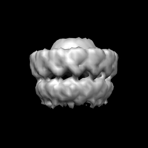
- EMDB-2136: Negative stain EM composite structure (part 1) of the type IV sec... -
+ Open data

- Basic information
| Entry | Database: EMDB / ID: EMD-2136 | |||||||||
|---|---|---|---|---|---|---|---|---|---|---|
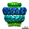
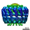
| Title | Negative stain EM composite structure (part 1) of the type IV secretion system subcomplex VirB4-VirB7-VirB9-VirB10 Secretion Secretion | |||||||||
 Map data Map data | Core complex part of the VirB4-core complex composite reconstruction | |||||||||
 Sample Sample |
| |||||||||
| Biological species |  Escherichia coli (E. coli) Escherichia coli (E. coli) | |||||||||
| Method | single particle reconstruction / negative staining / Resolution: 20.0 Å | |||||||||
 Authors Authors | Williams R / Wallden K / Yan J / Lian PW / Wang L / Thalassinos K / Orlova EV / Waksman G | |||||||||
 Citation Citation | Journal: Proc Natl Acad Sci U S A / Year: 2012 Title: Structure of the VirB4 ATPase, alone and bound to the core complex of a type IV secretion system. Authors: Karin Walldén / Robert Williams / Jun Yan / Pei W Lian / Luchun Wang / Konstantinos Thalassinos / Elena V Orlova / Gabriel Waksman /  Abstract: Type IV secretion (T4S) systems mediate the transfer of proteins and DNA across the cell envelope of bacteria. These systems play important roles in bacterial pathogenesis and in horizontal transfer ...Type IV secretion (T4S) systems mediate the transfer of proteins and DNA across the cell envelope of bacteria. These systems play important roles in bacterial pathogenesis and in horizontal transfer of antibiotic resistance. The VirB4 ATPase of the T4S system is essential for both the assembly of the system and substrate transfer. In this article, we present the crystal structure of the C-terminal domain of Thermoanaerobacter pseudethanolicus VirB4. This structure is strikingly similar to that of another T4S ATPase, VirD4, a protein that shares only 12% sequence identity with VirB4. The VirB4 domain purifies as a monomer, but the full-length protein is observed in a monomer-dimer equilibrium, even in the presence of nucleotides and DNAs. We also report the negative stain electron microscopy structure of the core complex of the T4S system of the Escherichia coli pKM101 plasmid, with VirB4 bound. In this structure, VirB4 is also monomeric and bound through its N-terminal domain to the core's VirB9 protein. Remarkably, VirB4 is observed bound to the side of the complex where it is ideally placed to play its known regulatory role in substrate transfer. | |||||||||
| History |
|
- Structure visualization
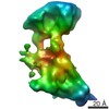
| Movie |
 Movie viewer Movie viewer |
|---|---|
| Structure viewer | EM map: SurfViewMolmilJmol/JSmol |
| Supplemental images |
- Downloads & links
-EMDB archive
| Map data | emd_2136.map.gz | 4.3 MB | EMDB map data format | |
|---|---|---|---|---|
| Header (meta data) | emd-2136-v30.xmlemd-2136.xml | 11.5 KB 11.5 KB | Display Display | EMDB header |
| Images | EMD2136.tif | 74.2 KB | ||
| Archive directory |  http://ftp.pdbj.org/pub/emdb/structures/EMD-2136ftp://ftp.pdbj.org/pub/emdb/structures/EMD-2136 http://ftp.pdbj.org/pub/emdb/structures/EMD-2136ftp://ftp.pdbj.org/pub/emdb/structures/EMD-2136 | HTTPS FTP |
-Related structure data
-Links
| EMDB pages | EMDB (EBI/PDBe) / EMDataResource |
|---|
-Map
| File | Download / File: emd_2136.map.gz / Format: CCP4 / Size: 58.2 MB / Type: IMAGE STORED AS FLOATING POINT NUMBER (4 BYTES) | ||||||||||||||||||||||||||||||||||||||||||||||||||||||||||||||||||||
|---|---|---|---|---|---|---|---|---|---|---|---|---|---|---|---|---|---|---|---|---|---|---|---|---|---|---|---|---|---|---|---|---|---|---|---|---|---|---|---|---|---|---|---|---|---|---|---|---|---|---|---|---|---|---|---|---|---|---|---|---|---|---|---|---|---|---|---|---|---|
| Annotation | Core complex part of the VirB4-core complex composite reconstruction | ||||||||||||||||||||||||||||||||||||||||||||||||||||||||||||||||||||
| Voxel size | X=Y=Z: 1.67 Å | ||||||||||||||||||||||||||||||||||||||||||||||||||||||||||||||||||||
| Density |
| ||||||||||||||||||||||||||||||||||||||||||||||||||||||||||||||||||||
| Symmetry | Space group: 1 | ||||||||||||||||||||||||||||||||||||||||||||||||||||||||||||||||||||
| Details | EMDB XML:
CCP4 map header:
| ||||||||||||||||||||||||||||||||||||||||||||||||||||||||||||||||||||
-Supplemental data
- Sample components
-Entire : TraB/TraN/TraO/TraF complex encoded by pKM101
| Entire | Name: TraB/TraN/TraO/TraF complex encoded by pKM101 |
|---|---|
| Components |
|
-Supramolecule #1000: TraB/TraN/TraO/TraF complex encoded by pKM101
| Supramolecule | Name: TraB/TraN/TraO/TraF complex encoded by pKM101 / type: sample / ID: 1000 Oligomeric state: 14-mer of core complex (TraN/TraO/TraF) and monomer of TraB Number unique components: 4 |
|---|---|
| Molecular weight | Experimental: 1.15 MDa / Theoretical: 1.15 MDa |
-Macromolecule #1: TraB
| Macromolecule | Name: TraB / type: protein_or_peptide / ID: 1 / Name.synonym: VirB4 Details: The 1.05 MDa core complex (TraN/TraO/TraF) with TraB bound, see EMD-2137 Number of copies: 1 / Recombinant expression: Yes |
|---|---|
| Source (natural) | Organism: Escherichia coli (E. coli) / Strain: pKM101 plasmid / Location in cell: cell envelope |
| Recombinant expression | Organism: Escherichia coli (E. coli) / Recombinant plasmid: pASK IBA3C |
-Macromolecule #2: TraN
| Macromolecule | Name: TraN / type: protein_or_peptide / ID: 2 / Name.synonym: VirB7 Details: The 1.05 MDa core complex (TraN/TraO/TraF) with TraB bound, see EMD-2137 Number of copies: 14 / Recombinant expression: Yes |
|---|---|
| Source (natural) | Organism: Escherichia coli (E. coli) / Strain: pKM101 plasmid / Location in cell: cell envelope |
| Recombinant expression | Organism: Escherichia coli (E. coli) / Recombinant plasmid: pASK IBA3C |
-Macromolecule #3: TraO
| Macromolecule | Name: TraO / type: protein_or_peptide / ID: 3 / Name.synonym: VirB9 Details: The 1.05 MDa core complex (TraN/TraO/TraF) with TraB bound, see EMD-2137 Number of copies: 14 / Recombinant expression: Yes |
|---|---|
| Source (natural) | Organism: Escherichia coli (E. coli) / Strain: pKM101 plasmid / Location in cell: cell envelope |
| Recombinant expression | Organism: Escherichia coli (E. coli) / Recombinant plasmid: pASK IBA3C |
-Macromolecule #4: TraF
| Macromolecule | Name: TraF / type: protein_or_peptide / ID: 4 / Name.synonym: VirB10 Details: The 1.05 MDa core complex (TraN/TraO/TraF) with TraB bound, see EMD-2137 Number of copies: 14 / Recombinant expression: Yes |
|---|---|
| Source (natural) | Organism: Escherichia coli (E. coli) / Strain: pKM101 plasmid / Location in cell: cell envelope |
| Recombinant expression | Organism: Escherichia coli (E. coli) / Recombinant plasmid: pASK IBA3C |
-Experimental details
-Structure determination
| Method | negative staining |
|---|---|
 Processing Processing | single particle reconstruction |
| Aggregation state | particle |
-Sample preparation
| Staining | Type: NEGATIVE / Details: NanoW |
|---|---|
| Grid | Details: Glow-discharged continuous carbon |
| Vitrification | Cryogen name: NONE / Instrument: OTHER |
- Electron microscopy
| Microscope | FEI TECNAI 12 |
|---|---|
| Electron beam | Acceleration voltage: 120 kV / Electron source: OTHER |
| Electron optics | Illumination mode: SPOT SCAN / Imaging mode: BRIGHT FIELDBright-field microscopy / Nominal defocus max: 2.0 µm / Nominal defocus min: 0.7 µm / Nominal magnification: 42000 |
| Sample stage | Specimen holder model: SIDE ENTRY, EUCENTRIC |
| Date | Dec 22, 2009 |
| Image recording | Category: FILM / Film or detector model: GENERIC FILM / Digitization - Scanner: ZEISS SCAI / Digitization - Sampling interval: 7 µm / Number real images: 12 / Bits/pixel: 8 |
| Tilt angle min | 0 |
| Tilt angle max | 0 |
-Image processing
| Final two d classification | Number classes: 330 |
|---|---|
| Final reconstruction | Applied symmetry - Point group: C1 (asymmetric) / Algorithm: OTHER / Resolution.type: BY AUTHOR / Resolution: 20.0 Å / Resolution method: FSC 0.5 CUT-OFF / Software - Name: IMAGIC Details: This map is part of a composite structure of two maps. Number images used: 10000 |
