Movie
Movie Controller

[English] 日本語
 Yorodumi
- EMDB-2098: Cryo-electron microscopy reconstruction of doublecortin-stabilise... -
+ Open data

- Basic information
| Entry | Database: EMDB / ID: EMD-2098 | |||||||||
|---|---|---|---|---|---|---|---|---|---|---|
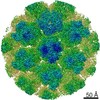
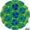
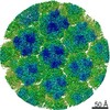
| Title | Cryo-electron microscopy reconstruction of doublecortin-stabilised microtubules in presence of kinesin | |||||||||
 Map data Map data | Reconstruction of doublecortin-stabilised microtubules decorated with kinesin motor domain (rigor) | |||||||||
 Sample Sample |
| |||||||||
 Keywords Keywords |  doublecortin / kinesin / microtubule / Microtubule-Associated Protein doublecortin / kinesin / microtubule / Microtubule-Associated Protein | |||||||||
| Function / homology |  Function and homology information Function and homology informationRHO GTPases activate KTN1 / Kinesins / COPI-dependent Golgi-to-ER retrograde traffic / membrane-bounded organelle / cytoplasm organization / plus-end-directed vesicle transport along microtubule / anterograde dendritic transport of neurotransmitter receptor complex / positive regulation of voltage-gated sodium channel activity / retrograde neuronal dense core vesicle transport / positive regulation of vesicle fusion ...RHO GTPases activate KTN1 / Kinesins / COPI-dependent Golgi-to-ER retrograde traffic / membrane-bounded organelle / cytoplasm organization / plus-end-directed vesicle transport along microtubule / anterograde dendritic transport of neurotransmitter receptor complex / positive regulation of voltage-gated sodium channel activity / retrograde neuronal dense core vesicle transport / positive regulation of vesicle fusion / anterograde axonal protein transport / MHC class II antigen presentation / cytoskeleton-dependent intracellular transport / vesicle transport along microtubule / positive regulation of intracellular protein transport / positive regulation of potassium ion transport / JUN kinase binding / plus-end-directed microtubule motor activity / stress granule disassembly / positive regulation of axon guidance / microtubule lateral binding / ciliary rootlet / centrosome localization / kinesin complex / synaptic vesicle transport / microtubule motor activity / microtubule-based movement / positive regulation of insulin secretion involved in cellular response to glucose stimulus / endocytic vesicle / centriolar satellite / microtubule-based process / phagocytic vesicle / axonal growth cone / axon cytoplasm / regulation of membrane potential / dendrite cytoplasm / axon guidance / positive regulation of synaptic transmission, GABAergic / hippocampus development / positive regulation of protein localization to plasma membrane / Hydrolases; Acting on acid anhydrides; Acting on GTP to facilitate cellular and subcellular movement / brain development / structural constituent of cytoskeleton / microtubule cytoskeleton organization / cellular response to type II interferon / microtubule cytoskeleton / mitotic cell cycle / nervous system development / microtubule binding / vesicle / microtubule / hydrolase activity / neuron projection / protein heterodimerization activity / GTPase activity / GTP binding / perinuclear region of cytoplasm / ATP hydrolysis activity / ATP binding / identical protein binding / metal ion binding / cytosol / cytoplasmSimilarity search - Function | |||||||||
| Biological species |  Bos taurus (cattle) / Bos taurus (cattle) /  Homo sapiens (human) / Rattus norvegicus (Norway rat) Homo sapiens (human) / Rattus norvegicus (Norway rat) | |||||||||
| Method | helical reconstruction / cryo EM / negative staining / Resolution: 8.2 Å | |||||||||
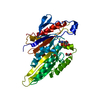
 Authors Authors | Liu JS / Schubert CR / Fu X / Fourniol FJ / Jaiswal JK / Houdusse A / Stultz CM / Moores CA / Walsh CA | |||||||||
 Citation Citation | Journal: J Cell Biol / Year: 2010 Title: Template-free 13-protofilament microtubule-MAP assembly visualized at 8 A resolution. Authors: Franck J Fourniol / Charles V Sindelar / Béatrice Amigues / Daniel K Clare / Geraint Thomas / Mylène Perderiset / Fiona Francis / Anne Houdusse / Carolyn A Moores /  Abstract: Microtubule-associated proteins (MAPs) are essential for regulating and organizing cellular microtubules (MTs). However, our mechanistic understanding of MAP function is limited by a lack of detailed ...Microtubule-associated proteins (MAPs) are essential for regulating and organizing cellular microtubules (MTs). However, our mechanistic understanding of MAP function is limited by a lack of detailed structural information. Using cryo-electron microscopy and single particle algorithms, we solved the 8 Å structure of doublecortin (DCX)-stabilized MTs. Because of DCX's unusual ability to specifically nucleate and stabilize 13-protofilament MTs, our reconstruction provides unprecedented insight into the structure of MTs with an in vivo architecture, and in the absence of a stabilizing drug. DCX specifically recognizes the corner of four tubulin dimers, a binding mode ideally suited to stabilizing both lateral and longitudinal lattice contacts. A striking consequence of this is that DCX does not bind the MT seam. DCX binding on the MT surface indirectly stabilizes conserved tubulin-tubulin lateral contacts in the MT lumen, operating independently of the nucleotide bound to tubulin. DCX's exquisite binding selectivity uncovers important insights into regulation of cellular MTs. | |||||||||
| History |
|
- Structure visualization
| Movie |
Movie viewer |
|---|---|
| Structure viewer | EM map: SurfViewMolmilJmol/JSmol |
| Supplemental images |

- Downloads & links
-EMDB archive
| Map data | emd_2098.map.gz | 1.5 MB | EMDB map data format | |
|---|---|---|---|---|
| Header (meta data) | emd-2098-v30.xmlemd-2098.xml | 15.9 KB 15.9 KB | Display Display | EMDB header |
| Images | 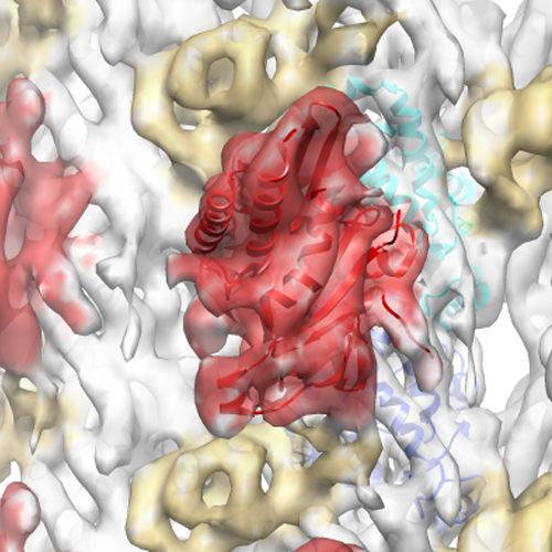 EMD-2098.jpg EMD-2098.jpg | 214.4 KB | ||
| Archive directory |  http://ftp.pdbj.org/pub/emdb/structures/EMD-2098ftp://ftp.pdbj.org/pub/emdb/structures/EMD-2098 http://ftp.pdbj.org/pub/emdb/structures/EMD-2098ftp://ftp.pdbj.org/pub/emdb/structures/EMD-2098 | HTTPS FTP |
-Related structure data
| Related structure data | 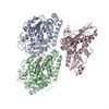 4atxMC  2095C 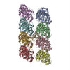 4atuC M: atomic model generated by this map C: citing same article ( |
|---|---|
| Similar structure data |


-Links
| EMDB pages | EMDB (EBI/PDBe) / EMDataResource |
|---|---|
| Related items in Molecule of the Month |


-Map
| File | Download / File: emd_2098.map.gz / Format: CCP4 / Size: 1.6 MB / Type: IMAGE STORED AS FLOATING POINT NUMBER (4 BYTES) | ||||||||||||||||||||||||||||||||||||||||||||||||||||||||||||||||||||
|---|---|---|---|---|---|---|---|---|---|---|---|---|---|---|---|---|---|---|---|---|---|---|---|---|---|---|---|---|---|---|---|---|---|---|---|---|---|---|---|---|---|---|---|---|---|---|---|---|---|---|---|---|---|---|---|---|---|---|---|---|---|---|---|---|---|---|---|---|---|
| Annotation | Reconstruction of doublecortin-stabilised microtubules decorated with kinesin motor domain (rigor) | ||||||||||||||||||||||||||||||||||||||||||||||||||||||||||||||||||||
| Voxel size | X=Y=Z: 2.8 Å | ||||||||||||||||||||||||||||||||||||||||||||||||||||||||||||||||||||
| Density |
| ||||||||||||||||||||||||||||||||||||||||||||||||||||||||||||||||||||
| Symmetry | Space group: 1 | ||||||||||||||||||||||||||||||||||||||||||||||||||||||||||||||||||||
| Details | EMDB XML:
CCP4 map header:
| ||||||||||||||||||||||||||||||||||||||||||||||||||||||||||||||||||||
-Supplemental data
- Sample components
-Entire : Doublecortin-stabilised microtubules decorated with kinesin motor...
| Entire | Name: Doublecortin-stabilised microtubules decorated with kinesin motor domains |
|---|---|
| Components |
|
-Supramolecule #1000: Doublecortin-stabilised microtubules decorated with kinesin motor...
| Supramolecule | Name: Doublecortin-stabilised microtubules decorated with kinesin motor domains type: sample / ID: 1000 / Oligomeric state: 13-protofilament microtubule / Number unique components: 4 |
|---|
-Macromolecule #1: Tubulin alpha-1D chain
| Macromolecule | Name: Tubulin alpha-1D chain / type: protein_or_peptide / ID: 1 / Oligomeric state: Heterodimer / Recombinant expression: No / Database: NCBI |
|---|---|
| Source (natural) | Organism: Bos taurus (cattle) / synonym: Cattle / Tissue: Brain / Location in cell: Cytoplasm |
| Molecular weight | Theoretical: 50 KDa |
| Sequence | InterPro: Alpha tubulin |
-Macromolecule #2: Doublecortin
| Macromolecule | Name: Doublecortin / type: protein_or_peptide / ID: 2 / Name.synonym: DCX / Recombinant expression: Yes |
|---|---|
| Source (natural) | Organism: Homo sapiens (human) / synonym: Human |
| Molecular weight | Theoretical: 40 KDa |
| Recombinant expression | Organism:   Spodoptera frugiperda (fall armyworm) / Recombinant plasmid: pFastBac Spodoptera frugiperda (fall armyworm) / Recombinant plasmid: pFastBac |
| Sequence | InterPro: Doublecortin domain |
-Macromolecule #3: Tubulin beta-2B chain
| Macromolecule | Name: Tubulin beta-2B chain / type: protein_or_peptide / ID: 3 / Oligomeric state: Heterodimer / Recombinant expression: No / Database: NCBI |
|---|---|
| Source (natural) | Organism: Bos taurus (cattle) / synonym: Cattle / Tissue: Brain / Location in cell: Cytoplasm |
| Molecular weight | Theoretical: 50 KDa |
| Sequence | InterPro: Beta tubulin, autoregulation binding site |
-Macromolecule #4: Kinesin motor domain
| Macromolecule | Name: Kinesin motor domain / type: protein_or_peptide / ID: 4 / Details: mutant T93N / Recombinant expression: Yes |
|---|---|
| Source (natural) | Organism: Rattus norvegicus (Norway rat) / synonym: Norway rat |
| Molecular weight | Theoretical: 40 KDa |
| Recombinant expression | Organism:  Escherichia coli (E. coli) Escherichia coli (E. coli) |
-Experimental details
-Structure determination
| Method | negative staining, cryo EM |
|---|---|
 Processing Processing | helical reconstruction |
| Aggregation state | filament |
-Sample preparation
| Buffer | pH: 6.8 Details: 20mM Pipes, 1mM EGTA, 3mM MgCl2, 1mM TCEP, 0.5mM GTP |
|---|---|
| Staining | Type: NEGATIVE / Details: cryo-EM |
| Grid | Details: 300 mesh lacey carbon grids |
| Vitrification | Cryogen name: ETHANE / Chamber humidity: 100 % / Instrument: FEI VITROBOT MARK I |
- Electron microscopy
| Microscope | FEI TECNAI F20 |
|---|---|
| Electron beam | Acceleration voltage: 200 kV / Electron source: FIELD EMISSION GUN |
| Electron optics | Illumination mode: FLOOD BEAM / Imaging mode: BRIGHT FIELDBright-field microscopy / Cs: 2.0 mm / Nominal defocus max: 2.9 µm / Nominal defocus min: 0.76 µm / Nominal magnification: 50000 |
| Sample stage | Specimen holder model: SIDE ENTRY, EUCENTRIC |
| Temperature | Average: 93 K |
| Alignment procedure | Legacy - Astigmatism: Objective lens astigmatism was corrected at 150,000 times magnification |
| Date | Sep 1, 2009 |
| Image recording | Category: FILM / Film or detector model: KODAK SO-163 FILM / Digitization - Scanner: ZEISS SCAI / Number real images: 63 / Average electron dose: 15 e/Å2 / Bits/pixel: 8 |
| Experimental equipment |  Model: Tecnai F20 / Image courtesy: FEI Company |
-Image processing
| CTF correction | Details: done with FREALIGN |
|---|---|
| Final reconstruction | Applied symmetry - Helical parameters - Δz: 9.2 Å Applied symmetry - Helical parameters - Δ&Phi: 27.7 ° Algorithm: OTHER / Resolution.type: BY AUTHOR / Resolution: 8.2 Å / Resolution method: FSC 0.5 CUT-OFF / Software - Name: SPIDER, FREALIGN Details: Approximately 168,000 asymmetric units were averaged together in the final map |
| Details | Particles were aligned using Spider and Frealign (Sindelar and Downing, 2010) |
-Atomic model buiding 1
| Initial model | PDB ID: Chain - #0 - Chain ID: E / Chain - #1 - Chain ID: F |
|---|---|
| Software | Name: Chimera |
| Details | Protocol: Rigid body |
| Refinement | Space: REAL / Protocol: RIGID BODY FIT / Target criteria: Cross correlation |
| Output model | PDB-4atx: |
-Atomic model buiding 2
| Initial model | PDB ID: Chain - Chain ID: A |
|---|---|
| Software | Name: Chimera, Flex-EM |
| Details | Protocol: Flexible fitting. Kinesin neck-linker (aa 324-329) was modeled into the EM density, and its position refined using Flex-EM, along with loop 2, loop 9 and helix alpha6 |
| Refinement | Space: REAL / Protocol: FLEXIBLE FIT / Target criteria: Cross correlation |
| Output model | PDB-4atx: |