 Movie
Movie Controller

[English] 日本語
 Yorodumi
- EMDB-2055: Acetylcholine-binding protein in the hemolymph of the planorbid s... -
+ Open data

- Basic information
| Entry | Database: EMDB / ID: EMD-2055 | |||||||||
|---|---|---|---|---|---|---|---|---|---|---|
| Title | Acetylcholine-binding protein in the hemolymph of the planorbid snail Biomphalaria glabrata is a pentagonal dodecahedron (60 subunits) | |||||||||
 Map data Map data | A negative temperature factor of 278.9 A^2 was applied to the map. | |||||||||
 Sample Sample |
| |||||||||
 Keywords Keywords |  ligand gated ion channel / LGIC / Cys-loop receptor / AChBP / acetylcholine binding protein / acetylcholine / AChR / acetylcholine receptor / Myasthenia gravis / pentagonal dodecahedron / nicotinic / dodecahedron / Schistosoma mansoni / bilharziosis / Biomphalaria glabrata / snail ligand gated ion channel / LGIC / Cys-loop receptor / AChBP / acetylcholine binding protein / acetylcholine / AChR / acetylcholine receptor / Myasthenia gravis / pentagonal dodecahedron / nicotinic / dodecahedron / Schistosoma mansoni / bilharziosis / Biomphalaria glabrata / snail | |||||||||
| Function / homology | extracellular ligand-gated monoatomic ion channel activity / Neurotransmitter-gated ion-channel / Neurotransmitter-gated ion-channel ligand-binding domain / Neurotransmitter-gated ion-channel ligand-binding domain superfamily / Neurotransmitter-gated ion-channel ligand binding domain / transmembrane signaling receptor activity / membrane => GO:0016020 / Acetylcholine-binding protein type 2 / Acetylcholine-binding protein type 1 Function and homology information Function and homology information | |||||||||
| Biological species |  Biomphalaria glabrata (bloodfluke planorb) Biomphalaria glabrata (bloodfluke planorb) | |||||||||
| Method | single particle reconstruction / cryo EM / Resolution: 6.0 Å | |||||||||
 Authors Authors | Saur M / Moeller V / Kapetanopoulos K / Braukmann S / Gebauer W / Tenzer S / Markl J | |||||||||
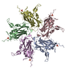
 Citation Citation | Journal: PLoS One / Year: 2012 Title: Acetylcholine-binding protein in the hemolymph of the planorbid snail Biomphalaria glabrata is a pentagonal dodecahedron (60 subunits). Authors: Michael Saur / Vanessa Moeller / Katharina Kapetanopoulos / Sandra Braukmann / Wolfgang Gebauer / Stefan Tenzer / Jürgen Markl /  Abstract: Nicotinic acetylcholine receptors (nAChR) play important neurophysiological roles and are of considerable medical relevance. They have been studied extensively, greatly facilitated by the gastropod ...Nicotinic acetylcholine receptors (nAChR) play important neurophysiological roles and are of considerable medical relevance. They have been studied extensively, greatly facilitated by the gastropod acetylcholine-binding proteins (AChBP) which represent soluble structural and functional homologues of the ligand-binding domain of nAChR. All these proteins are ring-like pentamers. Here we report that AChBP exists in the hemolymph of the planorbid snail Biomphalaria glabrata (vector of the schistosomiasis parasite) as a regular pentagonal dodecahedron, 22 nm in diameter (12 pentamers, 60 active sites). We sequenced and recombinantly expressed two ∼25 kDa polypeptides (BgAChBP1 and BgAChBP2) with a specific active site, N-glycan site and disulfide bridge variation. We also provide the exon/intron structures. Recombinant BgAChBP1 formed pentamers and dodecahedra, recombinant BgAChBP2 formed pentamers and probably disulfide-bridged di-pentamers, but not dodecahedra. Three-dimensional electron cryo-microscopy (3D-EM) yielded a 3D reconstruction of the dodecahedron with a resolution of 6 Å. Homology models of the pentamers docked to the 6 Å structure revealed opportunities for chemical bonding at the inter-pentamer interfaces. Definition of the ligand-binding pocket and the gating C-loop in the 6 Å structure suggests that 3D-EM might lead to the identification of functional states in the BgAChBP dodecahedron. | |||||||||
| History |
|
- Structure visualization
| Movie |
Movie viewer |
|---|---|
| Structure viewer | EM map: SurfViewMolmilJmol/JSmol |
| Supplemental images |

- Downloads & links
-EMDB archive
| Map data | emd_2055.map.gz | 83.7 MB | EMDB map data format | |
|---|---|---|---|---|
| Header (meta data) | emd-2055-v30.xmlemd-2055.xml | 12.5 KB 12.5 KB | Display Display | EMDB header |
| Images | 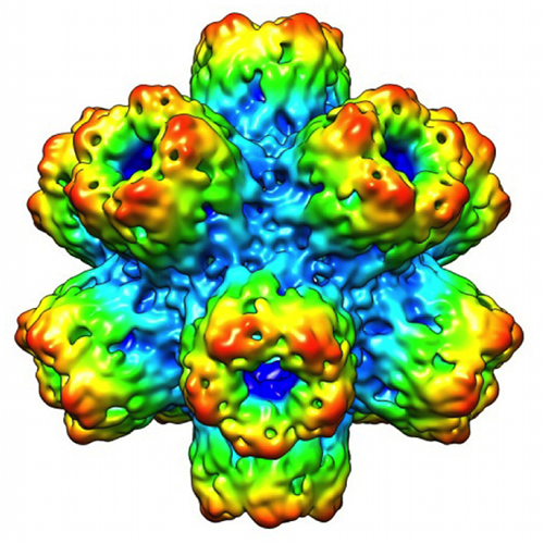 EMD-2055_gallery.png EMD-2055_gallery.png | 978.8 KB | ||
| Archive directory |  http://ftp.pdbj.org/pub/emdb/structures/EMD-2055ftp://ftp.pdbj.org/pub/emdb/structures/EMD-2055 http://ftp.pdbj.org/pub/emdb/structures/EMD-2055ftp://ftp.pdbj.org/pub/emdb/structures/EMD-2055 | HTTPS FTP |
-Related structure data
| Related structure data |  4aodMC  4aoeMC M: atomic model generated by this map C: citing same article ( |
|---|---|
| Similar structure data |


-Links
| EMDB pages | EMDB (EBI/PDBe) / EMDataResource |
|---|---|
| Related items in Molecule of the Month |


-Map
| File | Download / File: emd_2055.map.gz / Format: CCP4 / Size: 87.1 MB / Type: IMAGE STORED AS FLOATING POINT NUMBER (4 BYTES) | ||||||||||||||||||||||||||||||||||||||||||||||||||||||||||||||||||||
|---|---|---|---|---|---|---|---|---|---|---|---|---|---|---|---|---|---|---|---|---|---|---|---|---|---|---|---|---|---|---|---|---|---|---|---|---|---|---|---|---|---|---|---|---|---|---|---|---|---|---|---|---|---|---|---|---|---|---|---|---|---|---|---|---|---|---|---|---|---|
| Annotation | A negative temperature factor of 278.9 A^2 was applied to the map. | ||||||||||||||||||||||||||||||||||||||||||||||||||||||||||||||||||||
| Voxel size | X=Y=Z: 1 Å | ||||||||||||||||||||||||||||||||||||||||||||||||||||||||||||||||||||
| Density |
| ||||||||||||||||||||||||||||||||||||||||||||||||||||||||||||||||||||
| Symmetry | Space group: 1 | ||||||||||||||||||||||||||||||||||||||||||||||||||||||||||||||||||||
| Details | EMDB XML:
CCP4 map header:
| ||||||||||||||||||||||||||||||||||||||||||||||||||||||||||||||||||||
-Supplemental data
- Sample components
-Entire : Acetylcholine Binding Protein (AChBP) from Biomphalaria glabrata
| Entire | Name: Acetylcholine Binding Protein (AChBP) from Biomphalaria glabrata |
|---|---|
| Components |
|
-Supramolecule #1000: Acetylcholine Binding Protein (AChBP) from Biomphalaria glabrata
| Supramolecule | Name: Acetylcholine Binding Protein (AChBP) from Biomphalaria glabrata type: sample / ID: 1000 / Number unique components: 1 |
|---|---|
| Molecular weight | Experimental: 1.86 MDa / Theoretical: 1.86 MDa / Method: SDS-PAGE |
-Macromolecule #1: Biomphalaria glabrata Acetylcholine Binding Protein
| Macromolecule | Name: Biomphalaria glabrata Acetylcholine Binding Protein / type: protein_or_peptide / ID: 1 / Name.synonym: BgAChBP Details: As yet, it is not entirely clear whether the dodecahedron is a homomeric assembly of either type one and/or two, or a heteromeric assembly of types one and two. Number of copies: 60 / Oligomeric state: Dodecapentamer / Recombinant expression: No |
|---|---|
| Source (natural) | Organism: Biomphalaria glabrata (bloodfluke planorb) / Tissue: Hemolymph |
| Molecular weight | Experimental: 1.86 MDa / Theoretical: 1.86 MDa |
-Experimental details
-Structure determination
| Method | cryo EM |
|---|---|
 Processing Processing | single particle reconstruction |
| Aggregation state | particle |
-Sample preparation
| Concentration | 1.2 mg/mL |
|---|---|
| Buffer | pH: 7.4 / Details: 50 mM Tris-HCl, 5 mM MgCl2, 5mM CaCl2, 300mM NaCl |
| Grid | Details: C-flat holey carbon grids CF-2/2-3C-T, 30s glow discharge at 25 mA |
| Vitrification | Cryogen name: ETHANE / Chamber humidity: 97 % / Chamber temperature: 97 K / Instrument: GATAN CRYOPLUNGE 3 / Method: 2 x3.5 microlitres with 2 x 3s blotting |
- Electron microscopy #1
| Microscope | FEI TECNAI F20 |
|---|---|
| Electron beam | Acceleration voltage: 200 kV / Electron source: FIELD EMISSION GUN |
| Electron optics | Illumination mode: FLOOD BEAM / Imaging mode: BRIGHT FIELDBright-field microscopy / Cs: 2.0 mm / Nominal defocus max: 5.0 µm / Nominal defocus min: 2.0 µm / Nominal magnification: 50000 |
| Sample stage | Specimen holder model: GATAN LIQUID NITROGEN |
| Temperature | Average: 101 K |
| Microscopy ID | 1 |
| Alignment procedure | Legacy - Astigmatism: Objective lens astigmatism was corrected at 120,000 times magnification |
| Date | Jun 4, 2008 |
| Image recording | Category: FILM / Film or detector model: KODAK SO-163 FILM / Digitization - Scanner: PRIMESCAN / Digitization - Sampling interval: 5 µm / Number real images: 151 / Average electron dose: 30 e/Å2 / Bits/pixel: 8 |
| Tilt angle min | 0 |
| Tilt angle max | 0 |
| Experimental equipment |  Model: Tecnai F20 / Image courtesy: FEI Company |
-Electron microscopy #2
| Microscope | FEI TECNAI F20 |
|---|---|
| Electron beam | Acceleration voltage: 200 kV / Electron source: FIELD EMISSION GUN |
| Electron optics | Illumination mode: FLOOD BEAM / Imaging mode: BRIGHT FIELDBright-field microscopy / Cs: 2.0 mm / Nominal defocus max: 5.0 µm / Nominal defocus min: 2.0 µm / Nominal magnification: 50000 |
| Sample stage | Specimen holder model: GATAN LIQUID NITROGEN |
| Temperature | Average: 101 K |
| Microscopy ID | 2 |
| Alignment procedure | Legacy - Astigmatism: Objective lens astigmatism was corrected at 120,000 times magnification |
| Date | May 25, 2011 |
| Image recording | Category: FILM / Film or detector model: KODAK SO-163 FILM / Digitization - Scanner: PRIMESCAN / Digitization - Sampling interval: 5 µm / Number real images: 151 / Average electron dose: 30 e/Å2 / Bits/pixel: 8 |
| Tilt angle min | 0 |
| Tilt angle max | 0 |
| Experimental equipment | Model: Tecnai F20 / Image courtesy: FEI Company |
-Image processing
| CTF correction | Details: per micrograph |
|---|---|
| Final two d classification | Number classes: 608 |
| Final reconstruction | Applied symmetry - Point group: I (icosahedral) / Algorithm: OTHER / Resolution.type: BY AUTHOR / Resolution: 6.0 Å / Resolution method: FSC 0.5 CUT-OFF / Software - Name: EMAN1.9 / Number images used: 8374 |
| Details | manual selection with boxer, ctf-correction with FindCTF2d, refinement: EMAN1.9 (15 iterative cycles), final reconstruction using 1-degree references |
