Movie
Movie Controller

[English] 日本語
 Yorodumi
- EMDB-1932: Negative stain EM Map of the AAA protein CbbX, a red-type Rubisco... -
+ Open data

- Basic information
| Entry | Database: EMDB / ID: EMD-1932 | |||||||||
|---|---|---|---|---|---|---|---|---|---|---|
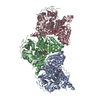
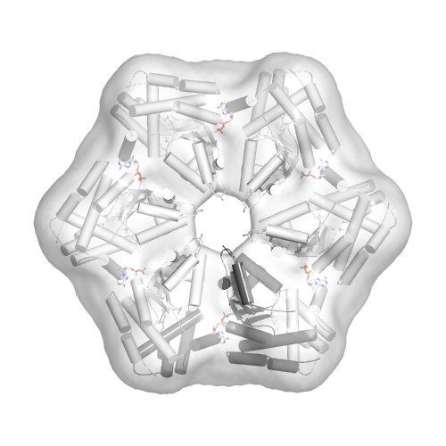
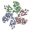
| Title | Negative stain EM Map of the AAA protein CbbX, a red-type Rubisco activase from R. sphaeroides | |||||||||
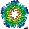
 Map data Map data | Negative stain EM reconstruction of the R. sphaeroides CbbX hexamer | |||||||||
 Sample Sample |
| |||||||||
 Keywords Keywords |  AAA+ protein / ATPase / Rubisco activase AAA+ protein / ATPase / Rubisco activase | |||||||||
| Function / homology |  Function and homology information Function and homology information | |||||||||
| Biological species |  Rhodobacter sphaeroides (bacteria) Rhodobacter sphaeroides (bacteria) | |||||||||
| Method | single particle reconstruction / negative staining / Resolution: 21.0 Å | |||||||||
 Authors Authors | Mueller-Cajar O / Stotz M / Wendler P / Hartl FU / Bracher A / Hayer-Hartl M | |||||||||
 Citation Citation | Journal: Nature / Year: 2011 Title: Structure and function of the AAA+ protein CbbX, a red-type Rubisco activase. Authors: Oliver Mueller-Cajar / Mathias Stotz / Petra Wendler / F Ulrich Hartl / Andreas Bracher / Manajit Hayer-Hartl /  Abstract: Ribulose 1,5-bisphosphate carboxylase/oxygenase (Rubisco) catalyses the fixation of atmospheric CO(2) in photosynthesis, but tends to form inactive complexes with its substrate ribulose 1,5- ...Ribulose 1,5-bisphosphate carboxylase/oxygenase (Rubisco) catalyses the fixation of atmospheric CO(2) in photosynthesis, but tends to form inactive complexes with its substrate ribulose 1,5-bisphosphate (RuBP). In plants, Rubisco is reactivated by the AAA(+) (ATPases associated with various cellular activities) protein Rubisco activase (Rca), but no such protein is known for the Rubisco of red algae. Here we identify the protein CbbX as an activase of red-type Rubisco. The 3.0-Å crystal structure of unassembled CbbX from Rhodobacter sphaeroides revealed an AAA(+) protein architecture. Electron microscopy and biochemical analysis showed that ATP and RuBP must bind to convert CbbX into functionally active, hexameric rings. The CbbX ATPase is strongly stimulated by RuBP and Rubisco. Mutational analysis suggests that CbbX functions by transiently pulling the carboxy-terminal peptide of the Rubisco large subunit into the hexamer pore, resulting in the release of the inhibitory RuBP. Understanding Rubisco activation may facilitate efforts to improve CO(2) uptake and biomass production by photosynthetic organisms. | |||||||||
| History |
|
- Structure visualization
| Movie |
Movie viewer |
|---|---|
| Structure viewer | EM map: SurfViewMolmilJmol/JSmol |
| Supplemental images |

- Downloads & links
-EMDB archive
| Map data | emd_1932.map.gz | 147 KB | EMDB map data format | |
|---|---|---|---|---|
| Header (meta data) | emd-1932-v30.xmlemd-1932.xml | 9.9 KB 9.9 KB | Display Display | EMDB header |
| Images |  emd-1932.png emd-1932.png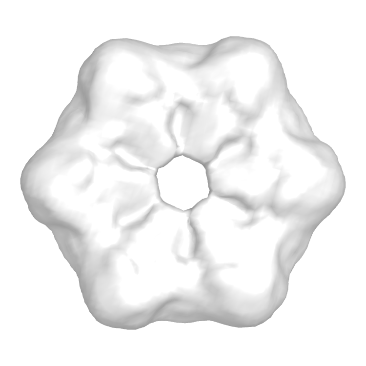 emd_1932.png emd_1932.png | 121.7 KB 105.4 KB | ||
| Archive directory |  http://ftp.pdbj.org/pub/emdb/structures/EMD-1932ftp://ftp.pdbj.org/pub/emdb/structures/EMD-1932 http://ftp.pdbj.org/pub/emdb/structures/EMD-1932ftp://ftp.pdbj.org/pub/emdb/structures/EMD-1932 | HTTPS FTP |
-Related structure data
| Related structure data | 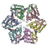 3zuhMC 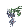 3sykC 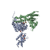 3sylC M: atomic model generated by this map C: citing same article ( |
|---|---|
| Similar structure data |

-Links
| EMDB pages | EMDB (EBI/PDBe) / EMDataResource |
|---|
-Map
| File | Download / File: emd_1932.map.gz / Format: CCP4 / Size: 7.8 MB / Type: IMAGE STORED AS FLOATING POINT NUMBER (4 BYTES) | ||||||||||||||||||||||||||||||||||||||||||||||||||||||||||||||||||||
|---|---|---|---|---|---|---|---|---|---|---|---|---|---|---|---|---|---|---|---|---|---|---|---|---|---|---|---|---|---|---|---|---|---|---|---|---|---|---|---|---|---|---|---|---|---|---|---|---|---|---|---|---|---|---|---|---|---|---|---|---|---|---|---|---|---|---|---|---|---|
| Annotation | Negative stain EM reconstruction of the R. sphaeroides CbbX hexamer | ||||||||||||||||||||||||||||||||||||||||||||||||||||||||||||||||||||
| Voxel size | X=Y=Z: 3.31 Å | ||||||||||||||||||||||||||||||||||||||||||||||||||||||||||||||||||||
| Density |
| ||||||||||||||||||||||||||||||||||||||||||||||||||||||||||||||||||||
| Symmetry | Space group: 1 | ||||||||||||||||||||||||||||||||||||||||||||||||||||||||||||||||||||
| Details | EMDB XML:
CCP4 map header:
| ||||||||||||||||||||||||||||||||||||||||||||||||||||||||||||||||||||
-Supplemental data
- Sample components
-Entire : R. sphaeroides CbbX 3D density map.
| Entire | Name: R. sphaeroides CbbX 3D density map. |
|---|---|
| Components |
|
-Supramolecule #1000: R. sphaeroides CbbX 3D density map.
| Supramolecule | Name: R. sphaeroides CbbX 3D density map. / type: sample / ID: 1000 / Oligomeric state: Hexamer / Number unique components: 1 |
|---|---|
| Molecular weight | Theoretical: 206 KDa |
-Macromolecule #1: Rubisco Activase
| Macromolecule | Name: Rubisco Activase / type: protein_or_peptide / ID: 1 / Name.synonym: CbbX Details: The protein is bound to Ribulose-1,5-bisphosphate, ATP and ATPgammaS Number of copies: 6 / Oligomeric state: Hexamer / Recombinant expression: Yes |
|---|---|
| Source (natural) | Organism: Rhodobacter sphaeroides (bacteria) |
| Molecular weight | Theoretical: 206 KDa |
| Recombinant expression | Organism: Escherichia coli BL21(DE3) (bacteria) / Recombinant plasmid: pHue |
| Sequence | GO: ATP binding |
-Experimental details
-Structure determination
| Method | negative staining |
|---|---|
 Processing Processing | single particle reconstruction |
| Aggregation state | particle |
-Sample preparation
| Concentration | 0.072 mg/mL |
|---|---|
| Buffer | pH: 8 Details: 20 mM Tris pH 8.0, 50 mM NaCl, 5mM MgCl2, 1mM Ribulose-1,5-bisphosphate, 1mM ATP/ATPgammaS |
| Staining | Type: NEGATIVE Details: Grids with adsorbed protein stained with 2% (w/v) uranyl acetate for 40 seconds. |
| Grid | Details: plain carbon grid |
| Vitrification | Cryogen name: NONE / Instrument: OTHER |
- Electron microscopy
| Microscope | FEI TECNAI 12 |
|---|---|
| Electron beam | Acceleration voltage: 120 kV / Electron source: LAB6 |
| Electron optics | Calibrated magnification: 90600 / Illumination mode: FLOOD BEAM / Imaging mode: BRIGHT FIELDBright-field microscopy / Cs: 2.0 mm / Nominal defocus max: 1.8 µm / Nominal defocus min: 0.26 µm / Nominal magnification: 90600 |
| Sample stage | Specimen holder: Eucentric / Specimen holder model: SIDE ENTRY, EUCENTRIC |
| Alignment procedure | Legacy - Astigmatism: objective lens astigmatism was corrected for at 110k magnification |
| Date | Nov 19, 2010 |
| Image recording | Category: CCD / Film or detector model: FEI EAGLE (2k x 2k) / Average electron dose: 20 e/Å2 |
-Image processing
| CTF correction | Details: phase flipping, each particle |
|---|---|
| Final reconstruction | Applied symmetry - Point group: C6 (6 fold cyclic) / Algorithm: OTHER / Resolution.type: BY AUTHOR / Resolution: 21.0 Å / Resolution method: FSC 0.5 CUT-OFF / Software - Name: MRC, IMAGIC, SPIDER / Number images used: 245 |
-Atomic model buiding 1
| Initial model | PDB ID: |
|---|---|
| Details | Protocol: rigid body |
| Refinement | Space: REAL / Protocol: RIGID BODY FIT |
| Output model | PDB-3zuh: |