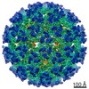
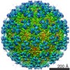
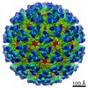
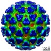
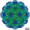
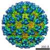
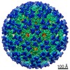
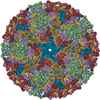
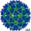
Journal: J Virol / Year: 2011 Title: The structure of barmah forest virus as revealed by cryo-electron microscopy at a 6-angstrom resolution has detailed transmembrane protein architecture and interactions. Authors: Victor A Kostyuchenko / Joanita Jakana / Xiangan Liu / Andrew D Haddow / Myint Aung / Scott C Weaver / Wah Chiu / Shee-Mei Lok / Abstract: Barmah Forest virus (BFV) is a mosquito-borne alphavirus that infects humans. A 6-Å-resolution cryo-electron microscopy three-dimensional structure of BFV exhibits a typical alphavirus organization, ...Barmah Forest virus (BFV) is a mosquito-borne alphavirus that infects humans. A 6-Å-resolution cryo-electron microscopy three-dimensional structure of BFV exhibits a typical alphavirus organization, with RNA-containing nucleocapsid surrounded by a bilipid membrane anchored with the surface proteins E1 and E2. The map allows details of the transmembrane regions of E1 and E2 to be seen. The C-terminal end of the E2 transmembrane helix binds to the capsid protein. Following the E2 transmembrane helix, a short α-helical endodomain lies on the inner surface of the lipid envelope. The E2 endodomain interacts with E1 transmembrane helix from a neighboring E1-E2 trimeric spike, thereby acting as a spacer and a linker between spikes. In agreement with previous mutagenesis studies, the endodomain plays an important role in recruiting other E1-E2 spikes to the budding site during virus assembly. The E2 endodomain may thus serve as a target for antiviral drug design.
History
Deposition
Mar 16, 2011
-
Header (metadata) release
Aug 12, 2011
-
Map release
Jan 27, 2012
-
Update
Oct 12, 2016
-
Current status
Oct 12, 2016
Processing site: PDBe / Status: Released
-
Structure visualization
Movie
Surface view with section colored by density value
Cryogen name: ETHANE / Chamber temperature: 100 K / Instrument: OTHER / Details: Vitrification instrument: Vitrobot / Method: Blot for 1 second before plunging
-
Electron microscopy
Microscope
JEOL 3200FSC
Electron beam
Acceleration voltage: 300 kV / Electron source: FIELD EMISSION GUN
In the structure databanks used in Yorodumi, some data are registered as the other names, "COVID-19 virus" and "2019-nCoV". Here are the details of the virus and the list of structure data.
Jan 31, 2019. EMDB accession codes are about to change! (news from PDBe EMDB page)
EMDB accession codes are about to change! (news from PDBe EMDB page)
The allocation of 4 digits for EMDB accession codes will soon come to an end. Whilst these codes will remain in use, new EMDB accession codes will include an additional digit and will expand incrementally as the available range of codes is exhausted. The current 4-digit format prefixed with “EMD-” (i.e. EMD-XXXX) will advance to a 5-digit format (i.e. EMD-XXXXX), and so on. It is currently estimated that the 4-digit codes will be depleted around Spring 2019, at which point the 5-digit format will come into force.
The EM Navigator/Yorodumi systems omit the EMD- prefix.
Related info.:Q: What is EMD? / ID/Accession-code notation in Yorodumi/EM Navigator
Yorodumi is a browser for structure data from EMDB, PDB, SASBDB, etc.
This page is also the successor to EM Navigator detail page, and also detail information page/front-end page for Omokage search.
The word "yorodu" (or yorozu) is an old Japanese word meaning "ten thousand". "mi" (miru) is to see.
Related info.:EMDB / PDB / SASBDB / Comparison of 3 databanks / Yorodumi Search / Aug 31, 2016. New EM Navigator & Yorodumi / Yorodumi Papers / Jmol/JSmol / Function and homology information / Changes in new EM Navigator and Yorodumi
 Movie
Movie Controller


 Open data
 Basic information
 Map data
 Sample
 Keywords
 Alphavirus /
  Function and homology information
 Authors
 Citation
 Structure visualization
 Downloads & links
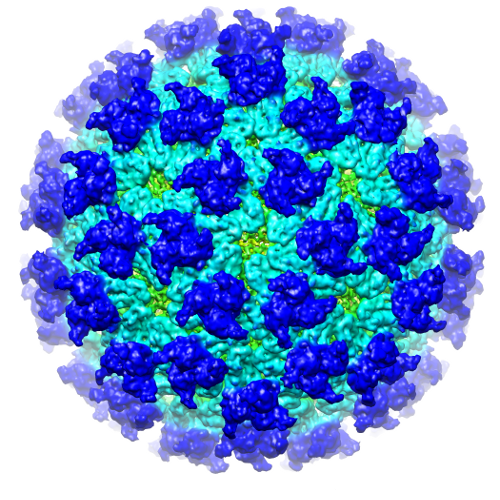 EMD-1886.png
 http://ftp.pdbj.org/pub/emdb/structures/EMD-1886
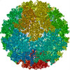


 Sample components
 Processing
 Electron microscopy