Movie
Movie Controller


+ Open data

- Basic information
| Entry | Database: EMDB / ID: EMD-1532 | |||||||||
|---|---|---|---|---|---|---|---|---|---|---|
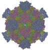
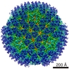
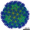
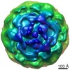
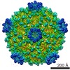
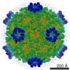
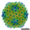
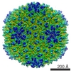
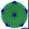
| Title | Three-dimensional structure of the Grass carp reovirus core. | |||||||||
 Map data Map data | Three-dimensional structure of the grass carp reovirus core. | |||||||||
 Sample Sample |
| |||||||||
 Keywords Keywords | Grass carp reovirus /  aquareovirus / dsRNA virus / 3D structure / reoviridae / cryo-electron microscopy aquareovirus / dsRNA virus / 3D structure / reoviridae / cryo-electron microscopy | |||||||||
| Biological species |  Grass carp reovirus Grass carp reovirus | |||||||||
| Method | single particle reconstruction / cryo EM / Resolution: 9.0 Å | |||||||||
 Authors Authors | Cheng L / Fang Q / Shah S / Atanasov IC / Zhou ZH | |||||||||
 Citation Citation | Journal: J Mol Biol / Year: 2008 Title: Subnanometer-resolution structures of the grass carp reovirus core and virion. Authors: Lingpeng Cheng / Qin Fang / Sanket Shah / Ivo C Atanasov / Z Hong Zhou /  Abstract: Grass carp reovirus (GCRV) is a member of the Aquareovirus genus of the family Reoviridae, a large family of double-stranded RNA (dsRNA) viruses infecting plants, insects, fishes and mammals. We ...Grass carp reovirus (GCRV) is a member of the Aquareovirus genus of the family Reoviridae, a large family of double-stranded RNA (dsRNA) viruses infecting plants, insects, fishes and mammals. We report the first subnanometer-resolution three-dimensional structures of both GCRV core and virion by cryoelectron microscopy. These structures have allowed the delineation of interactions among the over 1000 molecules in this enormous macromolecular machine and a detailed comparison with other dsRNA viruses at the secondary-structure level. The GCRV core structure shows that the inner proteins have strong structural similarities with those of orthoreoviruses even at the level of secondary-structure elements, indicating that the structures involved in viral dsRNA interaction and transcription are highly conserved. In contrast, the level of similarity in structures decreases in the proteins situated in the outer layers of the virion. The proteins involved in host recognition and attachment exhibit the least similarities to other members of Reoviridae. Furthermore, in GCRV, the RNA-translocating turrets are in an open state and lack a counterpart for the sigma1 protein situated on top of the close turrets observed in mammalian orthoreovirus. Interestingly, the distribution and the organization of GCRV core proteins resemble those of the cytoplasmic polyhedrosis virus, a cypovirus and the structurally simplest member of the Reoviridae family. Our results suggest that GCRV occupies a unique structure niche between the simpler cypoviruses and the considerably more complex mammalian orthoreovirus, thus providing an important model for understanding the structural and functional conservation and diversity of this enormous family of dsRNA viruses. | |||||||||
| History |
|
- Structure visualization
| Movie |
 Movie viewer Movie viewer |
|---|---|
| Structure viewer | EM map: SurfViewMolmilJmol/JSmol |
| Supplemental images |

- Downloads & links
-EMDB archive
| Map data | emd_1532.map.gz | 369.1 MB | EMDB map data format | |
|---|---|---|---|---|
| Header (meta data) | emd-1532-v30.xmlemd-1532.xml | 9 KB 9 KB | Display Display | EMDB header |
| Images | 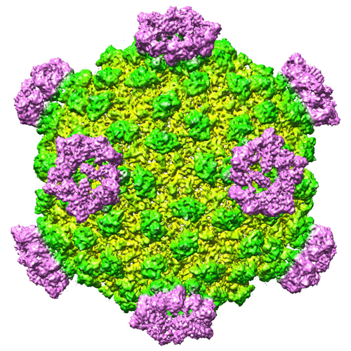 1532.gif 1532.gif | 152.2 KB | ||
| Archive directory |  http://ftp.pdbj.org/pub/emdb/structures/EMD-1532ftp://ftp.pdbj.org/pub/emdb/structures/EMD-1532 http://ftp.pdbj.org/pub/emdb/structures/EMD-1532ftp://ftp.pdbj.org/pub/emdb/structures/EMD-1532 | HTTPS FTP |
-Related structure data
-Links
| EMDB pages | EMDB (EBI/PDBe) / EMDataResource |
|---|
-Map
| File | Download / File: emd_1532.map.gz / Format: CCP4 / Size: 728.6 MB / Type: IMAGE STORED AS FLOATING POINT NUMBER (4 BYTES) | ||||||||||||||||||||||||||||||||||||||||||||||||||||||||||||||||||||
|---|---|---|---|---|---|---|---|---|---|---|---|---|---|---|---|---|---|---|---|---|---|---|---|---|---|---|---|---|---|---|---|---|---|---|---|---|---|---|---|---|---|---|---|---|---|---|---|---|---|---|---|---|---|---|---|---|---|---|---|---|---|---|---|---|---|---|---|---|---|
| Annotation | Three-dimensional structure of the grass carp reovirus core. | ||||||||||||||||||||||||||||||||||||||||||||||||||||||||||||||||||||
| Projections & slices | Image control
Images are generated by Spider. generated in cubic-lattice coordinate | ||||||||||||||||||||||||||||||||||||||||||||||||||||||||||||||||||||
| Voxel size | X=Y=Z: 1.16 Å | ||||||||||||||||||||||||||||||||||||||||||||||||||||||||||||||||||||
| Density |
| ||||||||||||||||||||||||||||||||||||||||||||||||||||||||||||||||||||
| Symmetry | Space group: 1 | ||||||||||||||||||||||||||||||||||||||||||||||||||||||||||||||||||||
| Details | EMDB XML:
CCP4 map header:
| ||||||||||||||||||||||||||||||||||||||||||||||||||||||||||||||||||||
 Z (Sec.)
 Y (Row.)
 X (Col.)


-Supplemental data
- Sample components
-Entire : Grass carp reovirus core
| Entire | Name: Grass carp reovirus core |
|---|---|
| Components |
|
-Supramolecule #1000: Grass carp reovirus core
| Supramolecule | Name: Grass carp reovirus core / type: sample / ID: 1000 / Number unique components: 5 |
|---|
-Supramolecule #1: Grass carp reovirus
| Supramolecule | Name: Grass carp reovirus / type: virus / ID: 1 / Name.synonym: aquareovirus / NCBI-ID: 128987 / Sci species name: Grass carp reovirus / Virus type: VIRION / Virus isolate: STRAIN / Virus enveloped: No / Virus empty: No / Syn species name: aquareovirus |
|---|---|
| Host (natural) | Organism:  Ctenopharyngodon idella (grass carp) / synonym: VERTEBRATES Ctenopharyngodon idella (grass carp) / synonym: VERTEBRATES |
| Virus shell | Shell ID: 1 / Name: GCRV core / Diameter: 810 Å / T number (triangulation number): 1 |
-Experimental details
-Structure determination
| Method | cryo EM |
|---|---|
 Processing Processing | single particle reconstruction |
| Aggregation state | particle |
-Sample preparation
| Concentration | 2.5 mg/mL |
|---|---|
| Buffer | pH: 7.5 Details: 10 mM PBS, 137mM NaCL, 2.7mM KCl, 8.1mM Na2HP04, 1.5mM KH2PO4 |
| Grid | Details: 400 mesh |
| Vitrification | Cryogen name: NITROGEN / Instrument: OTHER Method: Blot for 1 seconds before plunging. The sample was quickly plunged into a bath of liquid ethane cooled by liquid nitrogen. |
- Electron microscopy
| Microscope | FEI POLARA 300 |
|---|---|
| Electron beam | Acceleration voltage: 200 kV / Electron source: FIELD EMISSION GUN |
| Electron optics | Calibrated magnification: 129480 / Illumination mode: SPOT SCAN / Imaging mode: DIFFRACTION / Cs: 2.0 mm / Nominal defocus max: 2.0 µm / Nominal defocus min: 1.5 µm / Nominal magnification: 78000 |
| Sample stage | Specimen holder: Eucentric / Specimen holder model: GATAN LIQUID NITROGEN |
| Image recording | Category: CCD / Film or detector model: GENERIC TVIPS (4k x 4k) / Average electron dose: 20 e/Å2 / Bits/pixel: 32 |
| Experimental equipment |  Model: Tecnai Polara / Image courtesy: FEI Company |
-Image processing
| CTF correction | Details: Each microscoph |
|---|---|
| Final reconstruction | Applied symmetry - Point group: I (icosahedral) / Algorithm: OTHER / Resolution.type: BY AUTHOR / Resolution: 9.0 Å / Resolution method: FSC 0.5 CUT-OFF / Software - Name: IMIRS / Number images used: 3697 |
| Details | The particles were selected using an automatic selection program and verified manually. |
-Atomic model buiding 1
| Initial model | PDB ID: Chain - #0 - Chain ID: A / Chain - #1 - Chain ID: E |
|---|---|
| Software | Name: UCSF Chimera |
| Details | PDBEntryID_givenInChain. Protocol: Rigid Body. Automatically done by UCSF Chimera |
| Refinement | Space: REAL / Protocol: RIGID BODY FIT |